Radiological image detection apparatus and method of manufacturing the same

a technology of radiological image and detection apparatus, which is applied in the direction of radiation intensity measurement, x/gamma/cosmic radiation measurement, instruments, etc., can solve the problems of crystallinity disorder, mtf degradation, and clear increase of activator density, so as to improve mtf and increase the amount of luminescence

Inactive Publication Date: 2012-08-16
FUJIFILM CORP
View PDF4 Cites 10 Cited by
  • Summary
  • Abstract
  • Description
  • Claims
  • Application Information

AI Technical Summary

Benefits of technology

[0016]With the configuration and process, in the radiological image detection apparatus in which the first photodetector, the first scintillator, the second photodetector, and the second scintillator are arranged in order from the radiation incident side, a further increase of an amount of luminescence and a further improvement in MTF can be achieved.

Problems solved by technology

However, when such main luminescence region of the scintillator is examined in detail, the following problems still remain.
That is, an increase of an activator density poses clearly the technical problems mentioned hereunder.
In particular, when an activator density is enhanced in the initial phase of the vapor deposition of the scintillator, such enhancement has a tremendous adverse influence on the crystal growth of the scintillator, and the crystallinity is disordered.
Therefore, the lights are diffused between the columnar crystals, and thus degradation of MTF is caused.

Method used


Image

  • Radiological image detection apparatus and method of manufacturing the same
  • Radiological image detection apparatus and method of manufacturing the same
  • Radiological image detection apparatus and method of manufacturing the same

Examples


Embodiment Construction

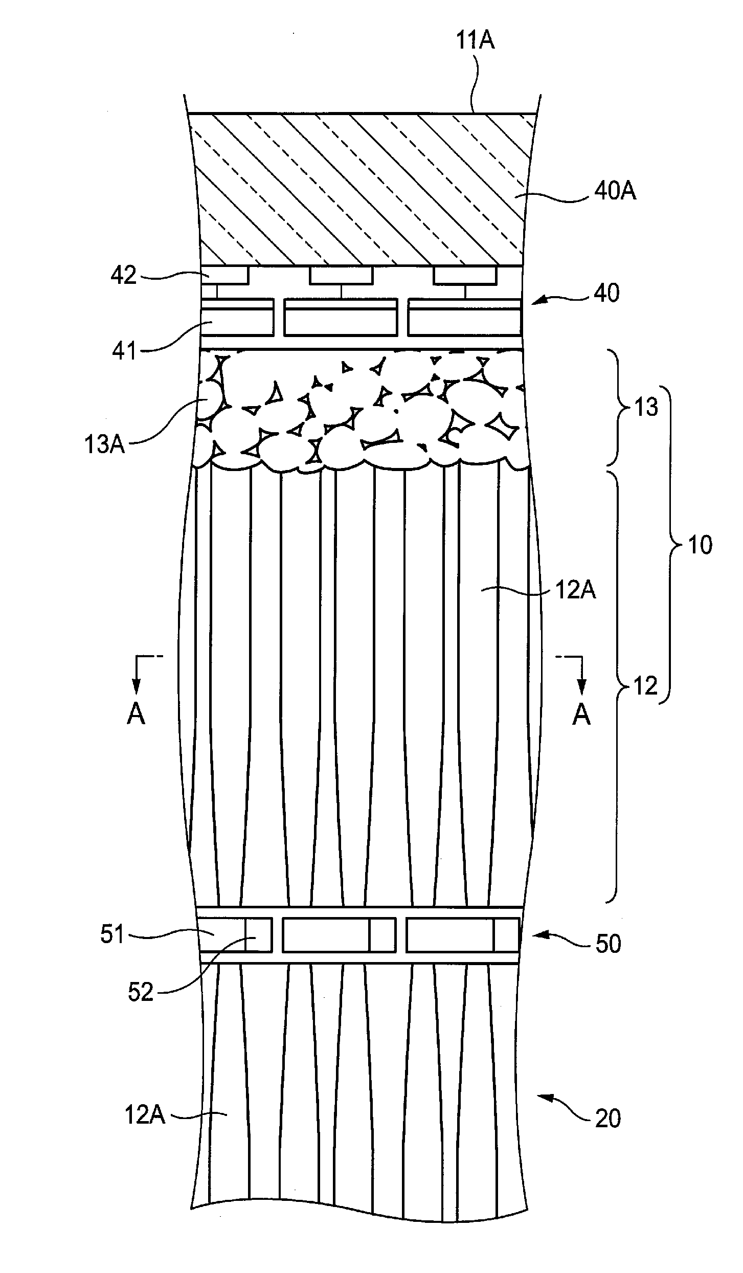
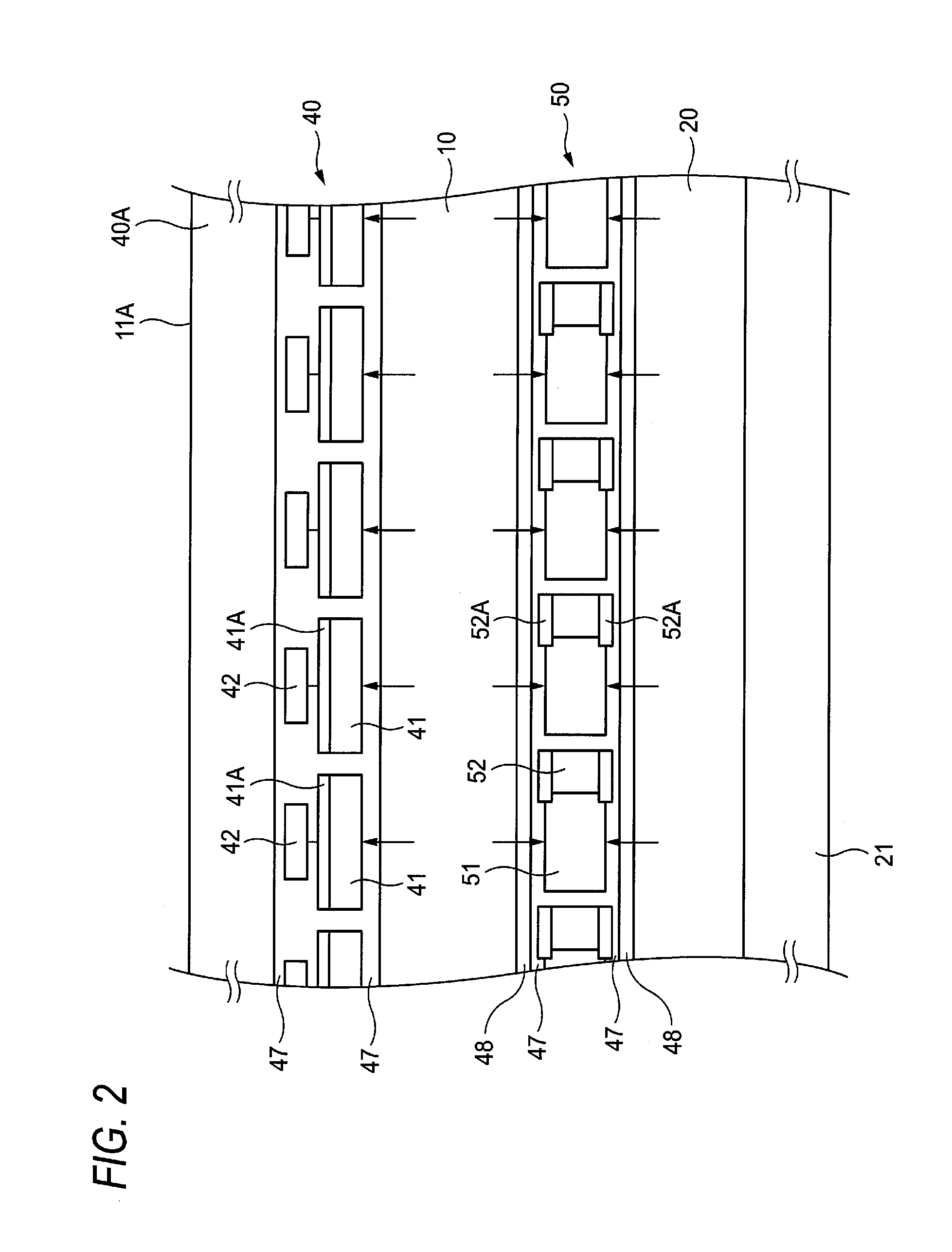
[0035]An example of an X-ray image detection apparatus (a radiological image detection apparatus) to explain an embodiment of the present invention will be explained with reference to FIG. 1 to FIG. 7B hereinafter.

[0036]Here, the same reference symbols are affixed to the similar configurations to those being already described, and their explanations will be omitted or simplified hereinafter.

[0037]In the following, explanation will be made by taking an X-ray image detection apparatus as one type of the radiological image detection apparatuses. A configuration described hereinafter is applicable to the radiological image detection apparatuses using various radiations such as α rays, β rays, γ rays, etc. According to these radiological image detection apparatuses using various radiations such as α rays, β rays, γ rays, etc., the operations and effects substantially similar to those described hereinafter can be achieved.

[1. Overall Configuration]

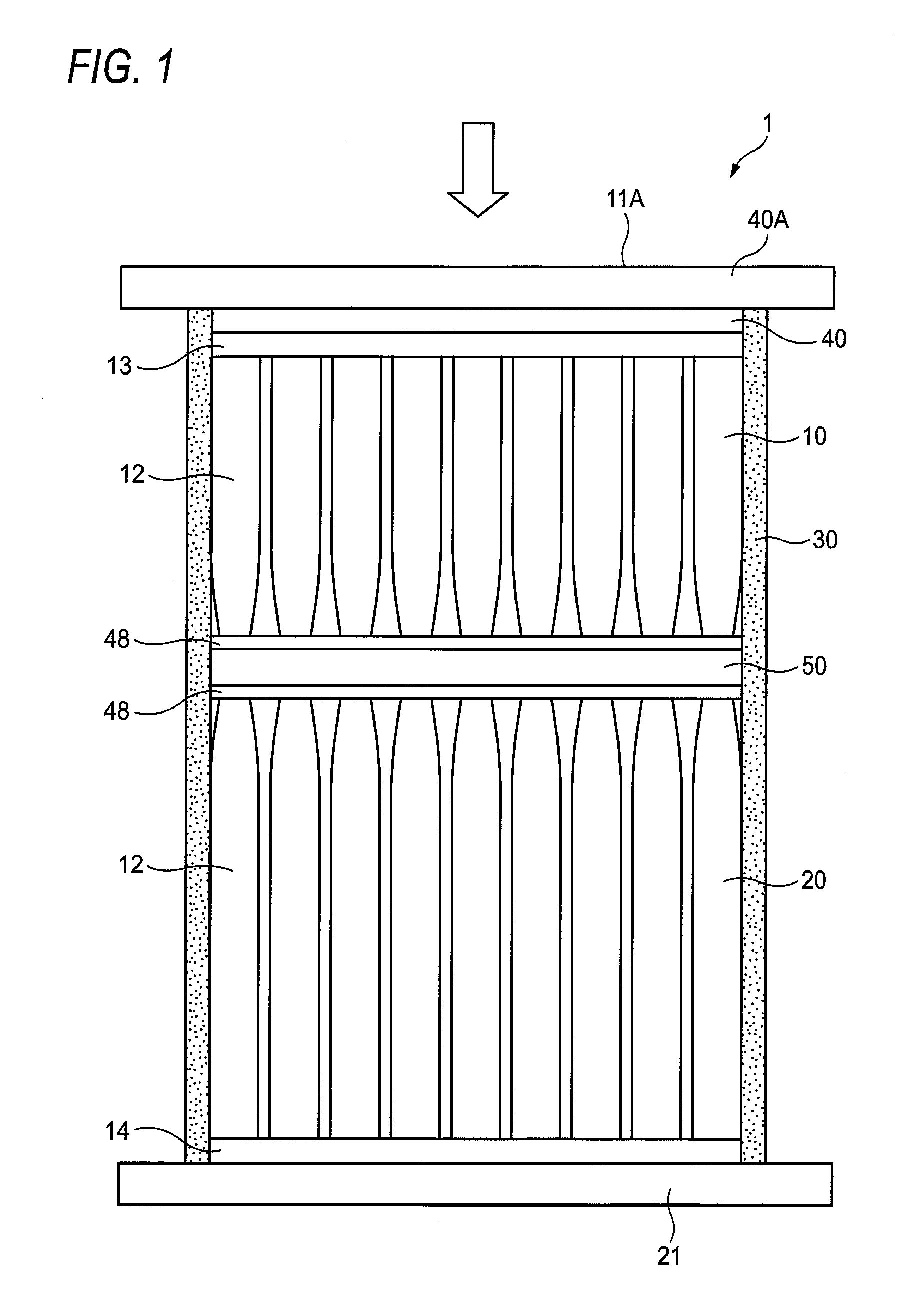
[0038]FIG. 1 is a side sectional view sho...


PUM


Abstract

A radiological image detection apparatus includes: a first scintillator and a second scintillator that emit fluorescent lights in response to irradiation of radiation; and a first photodetector and a second photodetector that detect the fluorescent lights; in which the first photodetector, the first scintillator, the second photodetector, and the second scintillator are arranged in order from a radiation incident side, and a high activator density region in which an activator density is relatively higher than an average activator density in a concerned scintillator is provided to at least one of the first scintillator located in vicinity of the first photodetector and the second scintillator located in vicinity of the second photodetector.

Description

CROSS-REFERENCE TO RELATED APPLICATIONS[0001]This application is based on and claims priority under 35 USC 119 from Japanese Patent Application No. 2011-028973 filed on Feb. 14, 2011; the entire content of which is incorporated herein by reference.BACKGROUND[0002]1. Technical Field[0003]The present invention relates to a radiological image detection apparatus used in the medical X-ray imaging system etc., and a method of manufacturing the same.[0004]2. Related Art[0005]In recent years, DR (Digital Radiography) using the X-ray image detection apparatus such as FPD (Flat Panel Detector) that converts an X-ray image into digital data, or the like is already put to practical use. In contrast to the former CR (Computed Radiography) system using the imaging plate formed of the stimulative phosphor (accumulative phosphor), this X-ray image detection apparatus has such a merit that the picked-up image can be checked on the spot, and thus its spread is proceeding apace.[0006]Various systems ...

Claims


Application Information

Patent Timeline
Patent Type & Authority Applications(United States)
IPC IPC(8): G01T1/202H01L31/18G01T1/20
CPCG01T1/242G01T1/2018H01L27/14663G01T1/20187G01T1/20189G01T1/20185G01T1/20181
Inventor NAKATSUGAWA, HARUYASUKANEKO, YASUHISAIWAKIRI, NAOTO
Owner FUJIFILM CORP
Why Eureka
Try Eureka