Method and system for expanding axial coverage in iterative reconstruction in computer tomography (CT)

a computer tomography and iterative reconstruction technology, applied in the field of image processing and system, can solve the problems of axial artifact reduction and iterative reconstruction algorithms failing to accurately reconstruct the region

US20130094735A1Active Publication Date: 2013-04-18TOSHIBA MEDICAL SYST CORP
4 Cites 19 Cited by

Patent Information

Authority / Receiving Office
US · United States
Patent Type
Applications(United States)
Current Assignee / Owner
Publication Date
2013-04-18

Smart Images

  • Figure 1
    Figure 1
  • Figure 2
    Figure 2
  • Figure 3
    Figure 3
Patent Text Reader

Abstract

The image generation method and system generates an image using a predetermined iterative reconstruction technique from cone beam data that has been expanded by adding additional data, and an instance of the iteration process is weighted according to a corresponding validation weight during the reconstruction. Optionally, an instance of the iteration process is weighted according to a combination of weights during the reconstruction. The predetermined combination of the weights includes axial weights based upon a validity value of the expanded data and statistical weights.
Need to check novelty before this filing date? Find Prior Art

Description

FIELD OF THE INVENTION

[0001] The current invention is generally related to an image processing and system, and more particularly related to the application of certain weights to a predetermined iterative reconstruction technique after the originally measured cone beam data has been expanded for substantially increasing the Z axial coverage.BACKGROUND OF THE INVENTION

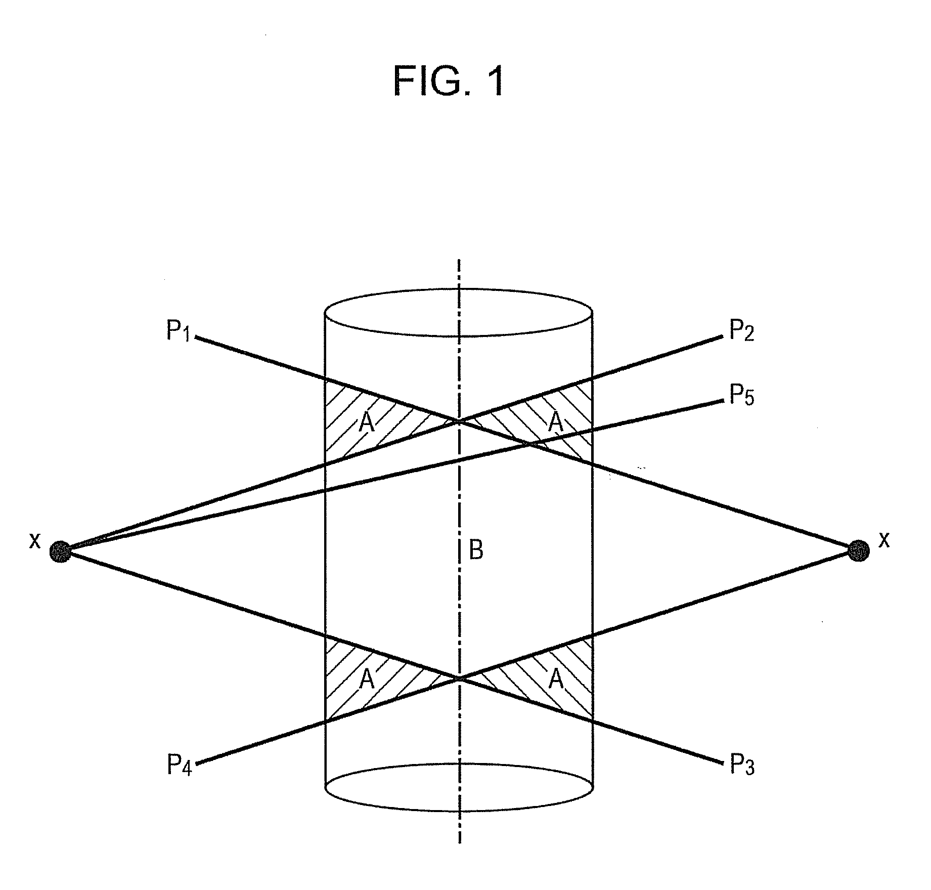
[0002] It is a well-known challenge in prior art to reconstruct an image of a long object along the z direction from circular cone beam data using an iterative reconstruction (IR) technique. The challenges involve the reduction in axial artifacts that IR suffers due to truncated measured data. For example, if an object to be imaged is confined within a support cylinder as shown in FIG. 1, projection data is acquired by use of the conventional circular cone-beam scanning geometry. The image volumes irradiated by x-rays are defined by the rays p1, p2, p3, and p4 as a source S is rotated around a predetermined vertical axis Z...

Examples

Embodiment Construction

)

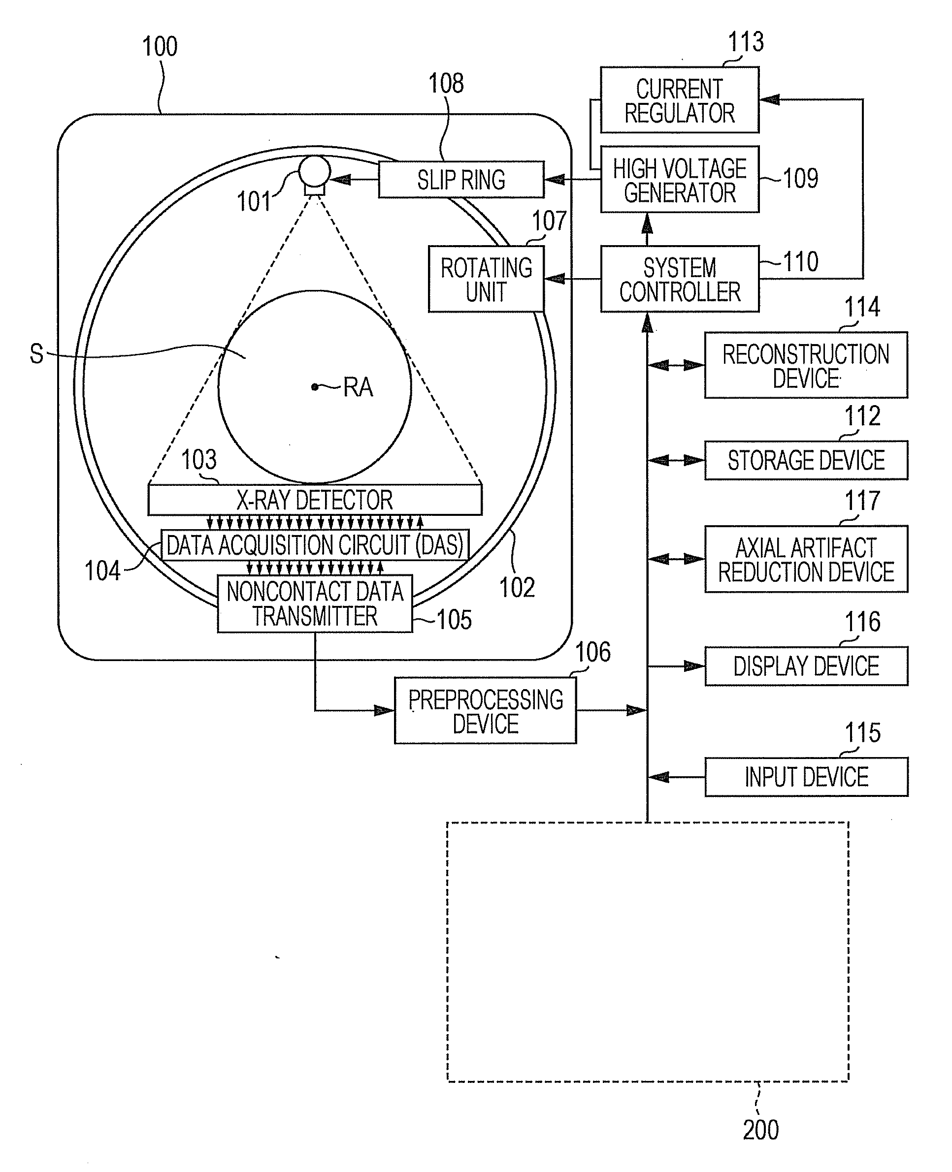
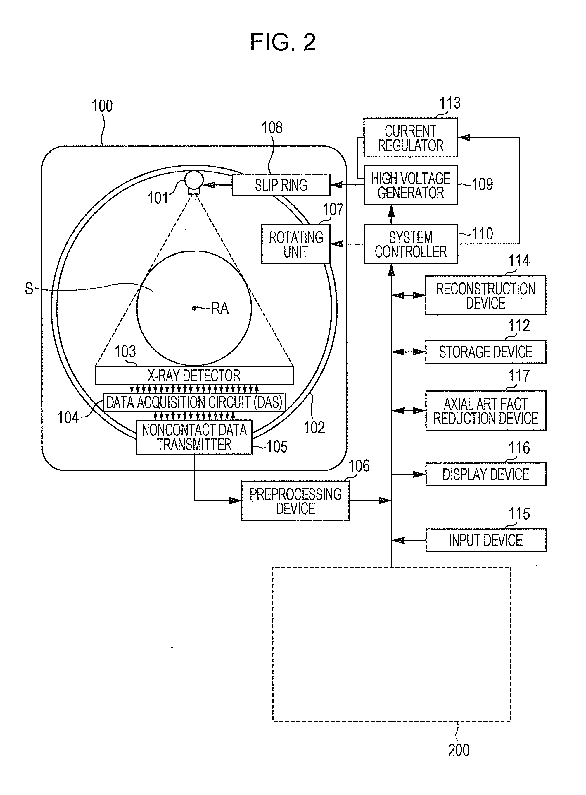
[0014]Referring now to the drawings, wherein like reference numerals designate corresponding structures throughout the views, and referring in particular to FIG. 2, a diagram illustrates one embodiment of the multi-slice X-ray CT apparatus or scanner according to the current invention including a gantry 100 and other devices or units. The gantry 100 is illustrated from a side view and further includes an X-ray tube 101, an annular frame 102 and a multi-row or two-dimensional array type X-ray detector 103. The X-ray tube 101 and X-ray detector 103 are diametrically mounted across a subject S on the annular frame 102, which is rotatably supported around a rotation axis RA. A rotating unit 107 rotates the frame 102 at a high speed such as 0.4 sec / rotation while the subject S is being moved along the axis RA into or out of the illustrated page.

[0015]The multi-slice X-ray CT apparatus further includes a high voltage generator 109 that applies a tube voltage to the X-ray tube 101 through...