
Antibody capable of recognizing HLA class i
a technology of antibodies and antibodies, applied in the field of antibodies capable of recognizing hla class i, can solve the problems of quick induction of cell death in human myeloma cells, and achieve the effect of prolonging the survival period of the mouse model and suppressing the cell growth of the im9 cell lin
Patent Information
- Authority / Receiving Office
- US · United States
- Patent Type
- Applications(United States)
- Current Assignee / Owner
- Publication Date
- 2012-09-27
- Estimated Expiration
- Not applicable · inactive patent
Smart Images
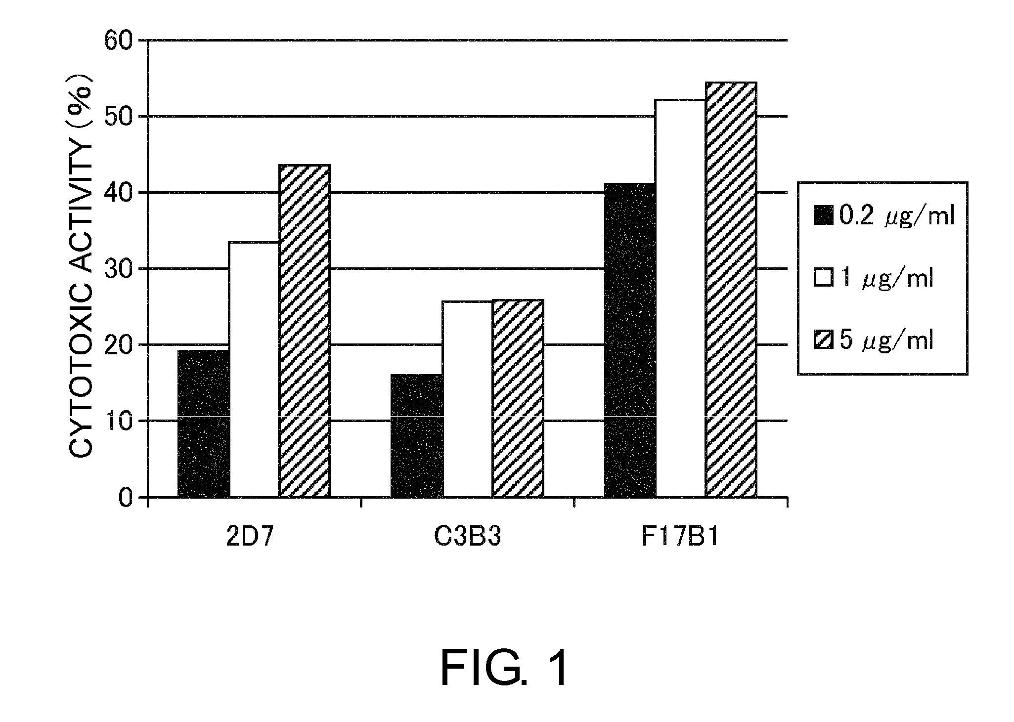
Figure 1 
Figure 2 
Figure 3
Abstract
Description
TECHNICAL FIELD
[0001] The present invention relates to HLA class I-recognizing antibodies.BACKGROUND ART
[0002] HLA, an important immune response molecule, is involved in recognizing and eliminating exogenous antigens, bacteria, virus-infected cells, and other such foreign substances. The main role of the HLA class I molecule is to present antigenic peptides (made up of about eight to ten amino acid residues) produced inside cells to CD8+ T cells. Accordingly, the HLA molecule plays a very important role in the immune response, and in immune tolerance induced by peptide presentation. HLA molecules are categorized into class I and class II. Class I molecules form a heterodimer made up of a 12-KD 132 microglobulin (β2M) and a 45-KD α-chain composed of three domains, α1-3. Class II molecules form a heterodimer made up of a 30-34 KD α-chain composed of two domains, α1 and α2, and a 26-29 KD β-chain composed of two domains, β1 and β2. HLA class I molecules are known to be further classified...
Examples
example 1
Preparation of Mouse anti-HLA class I Antibodies
Hybridoma Preparation
[0123]1.1. Preparation of Soluble HLA-A-FLAG (sHLA-A-flag) as Immunogen
1.1.1. Construction of an sHLA-A-flag Expression Vector and Establishment of an sHLA-A-flag-Producing CHO Line
[0124]sHLA-A-flag (SEQ ID NO: 1, a FLAG tag is located at positions 307 to 314), in which the flag tag is attached to the C terminus of the extracellular domain of HLA-A6802 (GenBank No. AM235885), was inserted into an animal cell expression vector. The nucleotide sequence was determined by a DNA sequencer, ABI PRISM 3700 DNA Sequencer (Applied Biosystems) using a BigDye Terminator Cycle Sequencing Kit (Applied Biosystems) according to the method described in the appended instruction manual. The constructed expression vector was introduced into CHO cells to establish an sHLA-A-flag-producing CHO line.
1.1.2. Purification of sHLA-A-flag
[0125]Using Q-Sepharose FF (GE Healthcare), ANTI-FLAG M2 Affinity Gel (SIGMA), and HiLoad 16 / 60 Superdex ...
example 2
Comparison of the Cytotoxic Activity of the F17B1, 2D7, and C3B3 Antibodies
[0129]The F17B1 antibody prepared as described in Example 1, and the 2D7 and C3B3 antibodies, which recognize HLA class I, were added to ARH77 cells at 0.2, 1, or 5 μg / ml. After the addition of antibodies, the cells were incubated at 37° C. for four to five hours. Then, the cells were stained with PI, and the proportion of nonviable cells was determined by FACS analysis (FIG. 1).
example 3
Determination of the Variable Regions of the Mouse anti-HLA class I Antibody F17B1
[0130]Total RNA was extracted from hybridomas using RNeasy Mini Kits (QIAGEN), and cDNA was synthesized using a SMART RACE cDNA Amplification Kit (BD Biosciences). Using PCR on the prepared cDNA, antibody variable region genes were inserted into cloning vectors. The nucleotide sequence of each DNA fragment was determined by a DNA sequencer, ABI PRISM 3700 DNA Sequencer (Applied Biosystems) using a BigDye Terminator Cycle Sequencing Kit (Applied Biosystems) according to the method described in the appended instruction manual. The determined H chain and L chain variable regions of the mouse F17B1 antibody are shown in SEQ ID NOs: 2 and 3, respectively. The CDRs and FRs were determined according to Kabat numbering.