Computer-assisted ultrasonic diagnosis method for left atrium/left auricle thrombus

A computer-aided ultrasonic diagnosis technology, applied in computing, special data processing applications, instruments, etc., can solve problems such as difficult identification of left atrial appendage thrombus images, deterioration of cardiac structure and function, failure to exclude thrombus in time, and avoid bleeding risks. Reduce the effect of cardiac structure and function, reduce the effect of subjective judgment

Active Publication Date: 2014-03-19
HARBIN MEDICAL UNIVERSITY
View PDF2 Cites 11 Cited by
  • Summary
  • Abstract
  • Description
  • Claims
  • Application Information

AI Technical Summary

Problems solved by technology

At present, transesophageal ultrasonography is used clinically. During this process, patients with atrial fibrillation will have obvious spontaneous development, which makes it difficult to distinguish the images of left atrial appendage thrombus and comb muscle. Therefore, suspected left atrium / left atrial appendage thrombus All patients were treated with warfarin (an anticoagulant) for four weeks, and then repeated transesophageal ultrasound to test the authenticity of suspected left atrial / LAA thrombus
On the one hand, this method makes patients who may not have thrombus receive unnecessary anticoagulant therapy, which increases the risk of bleeding (especially for elderly patients); on the other hand, for patients with thrombus, the existence of thrombus cannot be ruled out in time. Persistent atrial fibrillation worsens cardiac structure and function in patients with fibrillation

Method used


Image

  • Computer-assisted ultrasonic diagnosis method for left atrium/left auricle thrombus
  • Computer-assisted ultrasonic diagnosis method for left atrium/left auricle thrombus
  • Computer-assisted ultrasonic diagnosis method for left atrium/left auricle thrombus

Examples


Embodiment Construction

[0035] The following will clearly and completely describe the technical solutions in the embodiments of the present invention with reference to the accompanying drawings in the embodiments of the present invention. Obviously, the described embodiments are only part of the embodiments of the present invention, not all of them. Based on the embodiments of the present invention, all other embodiments obtained by persons of ordinary skill in the art without making creative efforts belong to the protection scope of the present invention.

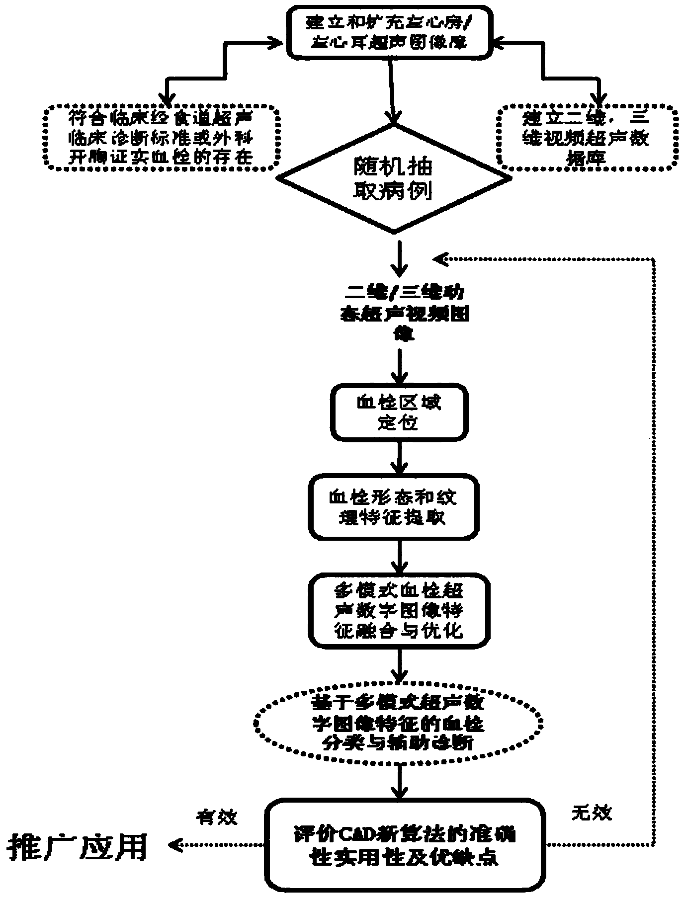
[0036] see figure 1 , figure 1 It is a schematic diagram of the system flow diagram of the invention, representing the general thinking of the present invention, and its specific steps are as follows:

[0037] Step 1: Based on actual clinical cases, we collect dynamic videos of thrombus from transesophageal multi-plane probes (according to the clinical diagnostic criteria of transesophageal ultrasound or the presence of thrombus confirmed by she...


PUM


Abstract

The invention discloses a computer-assisted ultrasonic diagnosis method for a left atrium/left auricle thrombus. The technical scheme includes that data mining technology, a pattern recognition theory and medical clinical information are combined, a gray-scale video and a real-time three-dimensional dynamic video serve as research objects, all-dimensional information in an image is accurately acquired, potential disease association rules in the image information are mined, and multiclass characteristics are comprehensively analyzed to obtain a detection method for automatically detecting and classifying the left atrium/left auricle thrombus. A thrombus recognition method can avoid missed diagnosis and misdiagnosis caused by subjective reasons such as inadequate experience or visual fatigue of doctors, and patients with suspected left atrium/left auricle thrombi clinically detected by transesophageal echocardiography can be confirmed as early as possible, so that the patients without thrombosis can receive cardioversion treatment of atrial fibrillation as early as possible. The method is simple and convenient to operate and high in practicability, and has important guiding significance for diagnosis and treatment of the left atrium/left auricle thrombus and ventricular fibrillation.

Description

technical field [0001] The invention relates to the technical field of computer-aided diagnosis, in particular to a method for computer-aided ultrasonic diagnosis of left atrium / left atrial appendage thrombus. Background technique [0002] Computer-aided diagnosis technology is referred to as CAD technology (Computer-Aided Diagnosis Technology). Computer-aided diagnosis includes at least two meanings: helping to find lesions and diagnosing lesions. The main advantage of computer-aided diagnosis of medical images lies in fast data processing and accurate quantitative calculation, which can provide clinical auxiliary diagnosis opinions with good consistency, high repeatability, objective and accurate, and reduce problems caused by doctors’ lack of experience or visual fatigue. Misdiagnosis caused by subjective reasons greatly expands the limited personal knowledge and experience of doctors, making the diagnosis more accurate and scientific. Therefore, although CAD is still i...

Claims


Application Information

Patent Timeline
Patent Type & Authority Applications(China)
IPC IPC(8): G06F17/50
Inventor 沈景霞李阳张英涛薛凤华程佳丽
Owner HARBIN MEDICAL UNIVERSITY
Why Eureka
Try Eureka