

Beagle spinal cord orientation channel stent and production method thereof
A beagle and spinal cord technology, applied in the field of beagle spinal cord directional channel support and its preparation based on three-dimensional printing technology, can solve problems such as spatial structure gap, and achieve the promotion of nerve regeneration, clear effect, strong self-renewal and proliferation ability Effect
- Summary
- Abstract
- Description
- Claims
- Application Information
AI Technical Summary
Problems solved by technology
Method used
Image

Examples
Embodiment 1
[0026] Embodiment 1 Beagle spinal cord directional channel support
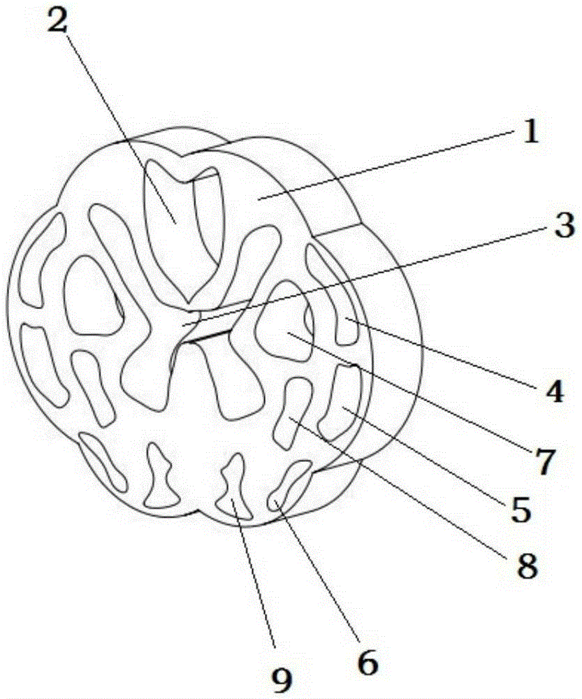
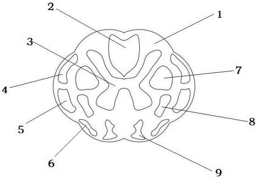
[0027] like figure 1 , 2 As shown, a Beagle spinal cord directional channel support includes a support body 1, a Beagle spinal cord directional channel support includes a support body 1, and the inside of the support body 1 is provided with thin bundles, wedge bundles, and spinothalamic bundles. , lateral corticospinal tract, posterior spinocerebellar tract, lateral spinothalamic tract, tectum of the spinal cord, anterior spinothalamic tract, anterior corticospinal tract, and the hollow area of gray matter; the hollow area 2 for growing thin and wedge tracts is located in the The upper part of the stent body 1; the hollow area 3 for growing gray matter is located at the center of the stent body 1; the hollow area 4 for growing the posterior spinocerebellar tract, the hollow area 5 for growing the lateral spinothalamus tract, There are two hollow areas 7 for growing the lateral corticospinal tract, two hol...
Embodiment 2
[0032] Embodiment 2 Preparation of Beagle Spinal Cord Oriented Channel Support
[0033] (1) According to the actual situation of the Beagle spinal cord, understand the diameter and course of the thin tracts, wedge tracts, corticospinal tracts, spinothalamic tracts, and spinocerebellar tracts, and design a 3D model of the Beagle spinal cord directional channel bracket, and the file is in STL format; Import the design model of the spinal cord directional channel bracket into the 3D bioprinter computer, and the printer software will convert the data of the model by itself;
[0034](2) In this embodiment, silk fibroin is used as the biological material of the spinal cord directional channel scaffold. Silk fibroin is prepared from silkworm cocoon silk by degumming, dissolving, dialysis and concentration methods. Put the silkworm cocoon silk at 80-120°C Na 2 CO 3 degumming in a solution of CaCl 2 , ethanol, and water solution for 2-5h, dialyzed in a dialysis bag under running wat...
Embodiment 3
[0038] Example Three Beagle Spinal Cord Oriented Channel Scaffold Preparation
[0039] (1) According to the actual situation of the Beagle spinal cord, understand the diameter and course of the thin tracts, wedge tracts, corticospinal tracts, spinothalamic tracts, and spinocerebellar tracts, and design a 3D model of the Beagle spinal cord directional channel bracket, and the file is in STL format; Import the design model of the spinal cord directional channel bracket into the 3D bioprinter computer, and the printer software will convert the data of the model by itself;
[0040] (2) In this embodiment, silk fibroin eggs and hydroxyapatite are used as the biomaterials of the spinal cord directional channel scaffolds, and are prepared by degumming, dissolving, dialysis and concentration methods, and silkworm cocoons are placed in 80-120°C Na 2 CO 3 degumming in a solution of CaCl 2 , ethanol, water (1:2:8) solution for 2-5h to dissolve, then dialyze in a dialysis bag under runn...
PUM
| Property | Measurement | Unit |
|---|---|---|
| Diameter | aaaaa | aaaaa |
| Wall thickness | aaaaa | aaaaa |
Abstract
Description
Claims
Application Information