Method, storage medium and device for extracting r-peak moment in electrocardiographic signal data

A technology of ECG signal and extraction method, applied in the field of signal processing, to achieve the effect of improving the success rate of defibrillation

Active Publication Date: 2022-03-04
鹤为科技(北京)有限公司
View PDF3 Cites 0 Cited by
  • Summary
  • Abstract
  • Description
  • Claims
  • Application Information

AI Technical Summary

Problems solved by technology

[0006] In view of this, the present invention provides a method, storage medium and device for extracting the R-peak moment in ECG signal data, so as to solve the problem of manual detection of ventricular fibrillation

Method used


Image

  • Method, storage medium and device for extracting r-peak moment in electrocardiographic signal data
  • Method, storage medium and device for extracting r-peak moment in electrocardiographic signal data
  • Method, storage medium and device for extracting r-peak moment in electrocardiographic signal data

Examples


Embodiment 1 Embodiment 2

[0064] Example as Figure 5 shown.

[0065] Step 201: sequentially extract the RR intervals in the ECG signal data, and store the newly extracted RR intervals into the newly recorded RR interval sequence;

[0066] Step 202: When the newly recorded RR interval sequence is not empty, take out the RR intervals in the newly recorded RR interval sequence sequentially, and when the RR intervals taken out continuously within the first preset duration are in the abnormal range, the probability is greater than the second preset value, execute step 203;

[0067] Step 203: Identify the waveform characteristics of the ECG signal data for the first preset time period. If the waveform characteristics conform to the preset oscillation state, execute step 103; otherwise, determine that no ventricular fibrillation is detected, and return to step 202.

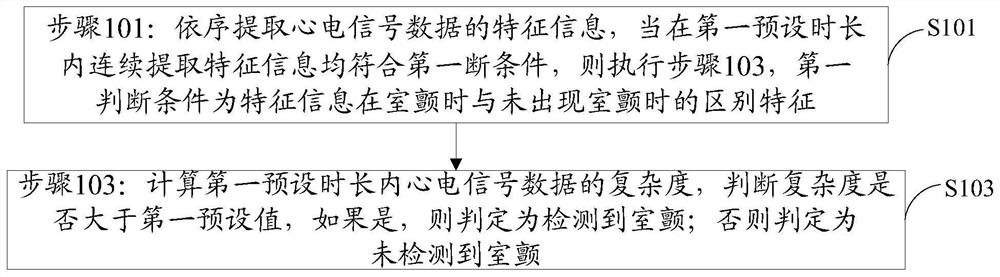
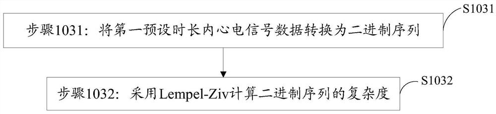
[0068] Step 103: Calculate the complexity of the ECG signal data for the first preset duration, and determine whether the complexity is great...

Embodiment 3

[0082] Embodiment three and embodiment four

[0083] Embodiment three such as Figure 7 shown.

[0084] Step 301: sequentially extract the waveform features in the ECG signal data, and store the newly extracted waveform features into the newly recorded waveform feature sequence;

[0085] Step 302: When the newly recorded waveform feature sequence is not empty, sequentially extract the waveform features in the newly recorded waveform feature sequence, and when the continuously extracted waveform features within the first preset duration conform to the preset oscillation state, then perform step 303;

[0086] Step 303: Extract the RR interval of the intracardiac signal data of the first preset time length, if the probability of the RR interval being in the abnormal range is greater than the second preset value, then execute step 103, otherwise it is determined that no ventricular fibrillation is detected, and return to step 302 .

[0087] Step 103: Calculate the complexity of...

Embodiment 4

[0088]Embodiment four such as Figure 8 shown.

[0089] Step 301-2: Sequentially extract the waveform features in the ECG signal data, store the newly extracted waveform features into the waveform feature sequence, and the newly extracted waveform features are unanalyzed waveform features in the waveform feature sequence;

[0090] Step 302-2: When the unanalyzed waveform features in the waveform feature sequence are not empty, read the unanalyzed waveform features sequentially, and convert the unanalyzed waveform features into analyzed waveform features after being read. If the waveform characteristics read continuously within the preset time period conform to the preset oscillation state, then step 303-2 is executed;

[0091] Step 303-2: Extract the RR interval of the ECG signal data of the first preset time length, if the probability of the RR interval being in the abnormal range is greater than the second preset value, then perform step 103, otherwise it is determined that...


PUM


Abstract

The present invention provides a method for extracting the R peak moment in electrocardiographic signal data, which reads the first N data of electrocardiographic signal data, which is recorded as data z(i), i=1, 2...N, and the N Each data contains at least one QRS wave, perform differential operation on z(i) to obtain the differential result dz(i), calculate the amplitude of z(i), and obtain the amplitude result Az(i); subdivide z(i) into K segment, T M (j) is the difference maximum value of the difference result dz(i) of the jth segment, T m (j) is the minimum difference value of the difference result dz(i) of the jth segment, T A (j) is the maximum amplitude value of the amplitude result Az(i) of the jth segment; update the maximum value of the difference, the minimum value of the difference and the maximum value of the amplitude to locate all that meet dz(i)≥T M The starting position of is the P1 sequence, positioning all dz(i)≤T m The starting position is the P2 sequence. The invention can accurately and quickly extract the R peak moment in the electrocardiographic signal data.

Description

technical field [0001] The invention relates to the field of signal processing, in particular to a method, storage medium and device for extracting R-peak time in electrocardiographic signal data. Background technique [0002] Ventricular fibrillation (VF, referred to as ventricular fibrillation) is a serious cardiovascular disease, which is caused by human factors such as coronary heart disease and myocardial infarction or external factors such as surgery and drug poisoning. When ventricular fibrillation occurs, the patient is usually unconscious, pulseless, and bloodless, and is at high risk, which may threaten the patient's life at any time. [0003] When a patient has ventricular fibrillation, timely detection and electric shock defibrillation are currently recognized as an important means of treatment for the patient. If defibrillation can be implemented within 1 minute of ventricular fibrillation, the success rate of defibrillation can be close to 100%. After 5 minute...

Claims


Application Information

Patent Timeline
Patent Type & Authority Patents(China)
IPC IPC(8): A61B5/318A61B5/352A61B5/366A61B5/353A61B5/355A61B5/36A61B5/361
CPCA61B5/361A61B5/318
Inventor 王金石
Owner 鹤为科技(北京)有限公司
Why Eureka
Try Eureka