Method and device for determining medical image splicing abnormity

A medical imaging and imaging technology, applied in the field of medical imaging, can solve the problems of incorrect image splicing, heavy workload of doctors and operators, and achieve the effect of improving efficiency and quality

Active Publication Date: 2021-10-22
浙江太美医疗科技股份有限公司
View PDF6 Cites 0 Cited by
  • Summary
  • Abstract
  • Description
  • Claims
  • Application Information

AI Technical Summary

Problems solved by technology

Most of the existing multi-bed image stitching methods are manually stitched or completed by scanning equipment. The workload of doctors and operators is heavy. During the stitching process, it is very likely that the wrong stitching of images is caused by not paying attention to the stitching position of the images.

Method used


Image

  • Method and device for determining medical image splicing abnormity
  • Method and device for determining medical image splicing abnormity
  • Method and device for determining medical image splicing abnormity

Examples


Embodiment Construction

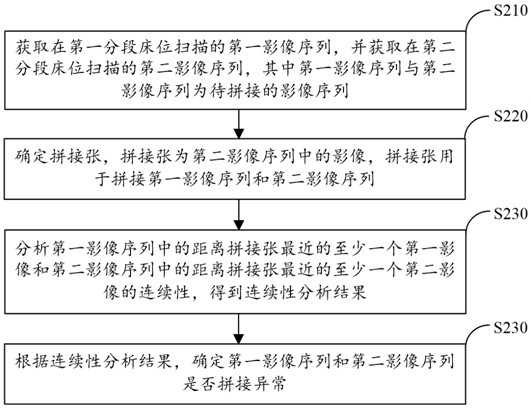
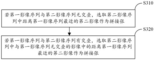
[0030] Embodiments of the present application will be described in more detail below with reference to the accompanying drawings. Although certain embodiments of the present application are shown in the drawings, it should be understood that the application may be embodied in various forms and should not be construed as limited to the embodiments set forth herein; A more thorough and complete understanding of the application. It should be understood that the drawings and embodiments of the present application are for exemplary purposes only, and are not intended to limit the protection scope of the present application.

[0031] The term "comprising" and its variants used in this application are open-ended, ie "including but not limited to". The term "based on" means "based on, at least in part." The term "one embodiment" means "at least one embodiment"; the term "another embodiment" means "at least one further embodiment". Relevant definitions of other terms will be given i...


PUM


Abstract

The invention provides a method for determining medical image splicing abnormity, and the method comprises the steps: obtaining a first image sequence scanned at a first sectional bed, and obtaining a second image sequence scanned at a second sectional bed, wherein the first image sequence and the second image sequence to be to-be-spliced image sequences; determining a spliced sheet, the spliced sheet being an image in the second image sequence, the spliced sheet being used for splicing the first image sequence and the second image sequence; analyzing the continuity of at least one first image closest to the spliced sheet in the first image sequence and at least one second image closest to the spliced sheet in the second image sequence to obtain a continuity analysis result; and determining whether splicing of the first image sequence and the second image sequence is abnormal or not according to a continuity analysis result. According to the method for determining the medical image splicing abnormity, by judging the continuity of the multi-bed spliced image, automatic reminding of the medical image splicing abnormity is achieved, doctors and operators are helped to quickly confirm the correctness of multi-bed image splicing, and the image processing efficiency and quality are improved.

Description

technical field [0001] The present application relates to the technical field of medical imaging, and more specifically, relates to a method and device for determining abnormality of medical image stitching. Background technique [0002] DICOM (Digital Imaging and Communications in Medicine) is an international standard (ISO 12052) for medical images and related information. It defines a medical image format that can be used for data exchange with a quality that meets clinical needs. [0003] In the process of human body image scanning, since the axial length of the human body is greater than the length of the scanner, it needs to be scanned bed by bed, so it is necessary to splicing the scanned images of multiple beds. Most of the existing multi-bed image stitching methods are manually stitched or completed by scanning equipment. The workload of doctors and operators is heavy. During the stitching process, it is very likely that the wrong stitching of images is caused by n...

Claims


Application Information

Patent Timeline
Patent Type & Authority Applications(China)
IPC IPC(8): G06T3/40G16H30/20
CPCG06T3/4038G06T3/4007G16H30/20
Inventor 潘伟凡
Owner 浙江太美医疗科技股份有限公司
Why Eureka
Try Eureka