
Quantitative multiplex methylation specific PCR method—cMethDNA, reagents, and its use
a methylation specific, quantitative technology, applied in the field of quantitative multiplex methylation specific pcr method, can solve the problems of lack of clinical validation studies, difficult reproducible detection of methylated dna from serum/plasma, lack of robustness of assay, etc., and achieve the effect of evaluating the response to treatmen
- Summary
- Abstract
- Description
- Claims
- Application Information
AI Technical Summary
Benefits of technology
Problems solved by technology
Method used
Image

Examples
example 1
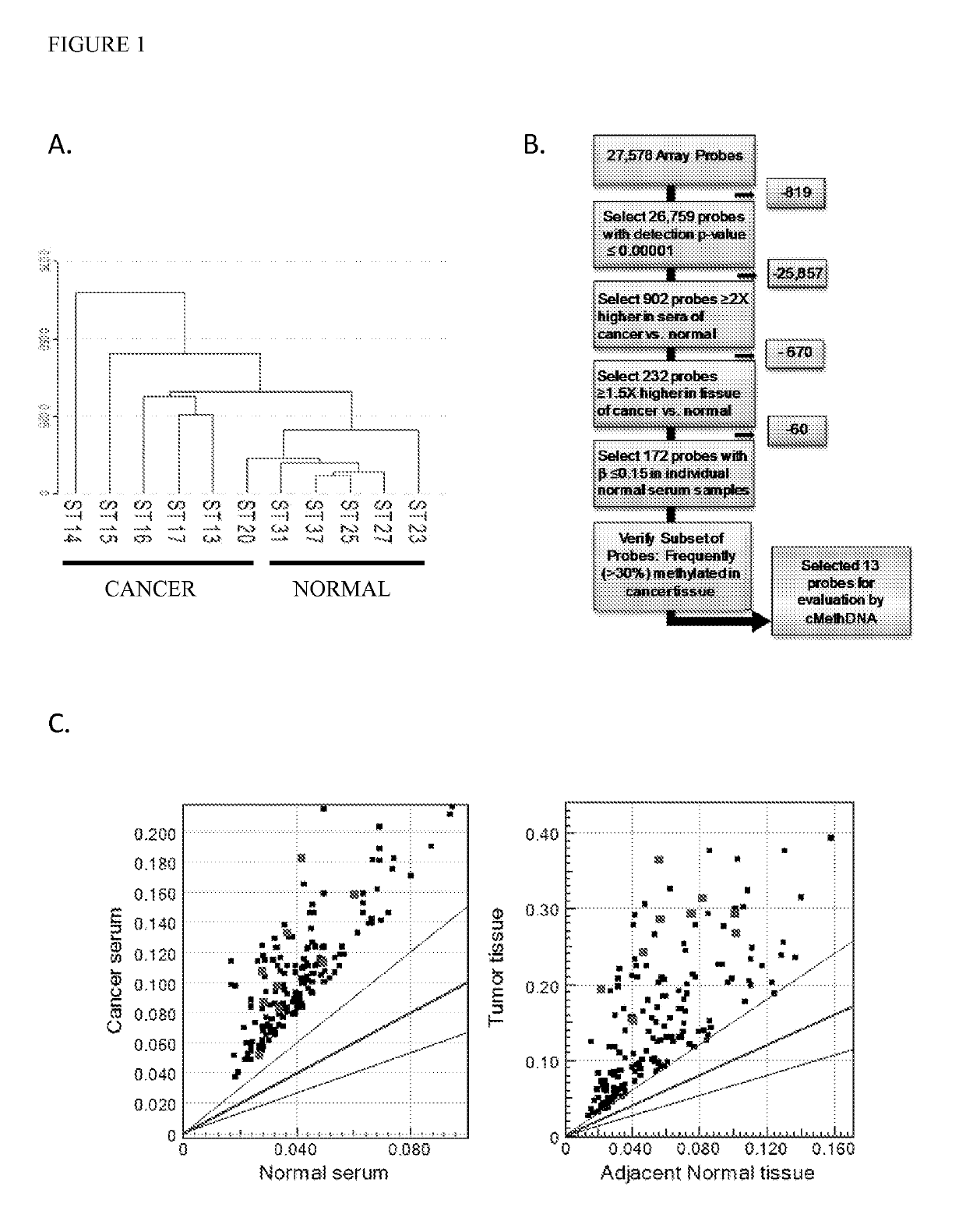
[0089]Recently, genome wide-DNA methylation arrays and bisulfite treated DNA sequencing of primary breast tumors have resulted in the identification of novel subgroups from the “intrinsic subtypes” identified by gene expression profiling (Breast Cancer Res. 2007; 9:R20; Pathobiology. 2008; 75:149-52; Anal Cell Pathol (Amst). 2010; 33:133-41; Cancer Epidemiol Biomarkers Prev. 2007; 16:1812-21; J Epidemiol. 2012; 22:384-94). As a first step to comprehensively analyze the methylation profile of circulating DNA in breast cancer patients, a methylation array analysis was conducted using the Infinium HumanMethylation27 BeadChip Array (Illumina, Inc.) on a discovery set of six recurrent metastatic breast cancer sera, five normal sera, and twenty normal leukocyte (four pools of 5 individuals each) (Table 1). An unsupervised hierarchical cluster analysis performed using 26759 probes (97% of the array; probe detection p-values <0.0001) found that cancer and normal sera clustered into two sepa...
example 2
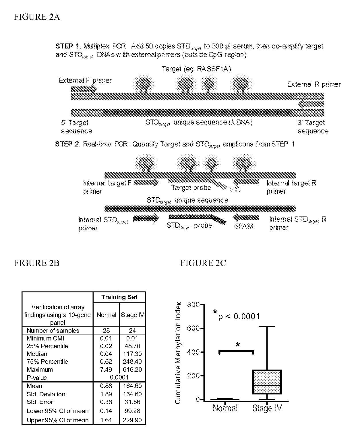
[0091]Marker detection frequency in circulating tumor DNA. In this example, fourteen genes hypermethylated in cancer sera compared to normal were tested-thirteen novel genes (HOXB4 miRNA, RASGRF2, ARHGEF7, AKR1B1, TMEFF2, TM6SF1, COL6A2, GPX7, HIST1H3C, ALX1, DAB1, DABIP2, and GAS7) identified by array analysis and RASSF1A, based upon our prior experience. Methylation frequency of the 14 genes was then verified in the patient and normal serum DNA samples used for arraying with the cMethDNA assay. The gene panel was refined by removing genes from the panel with high background or low frequency (FIG. 2B), specifically eliminating ALX1, DABIP2, and DAB1 which displayed levels of methylation of above 7 units (the set threshold for “normal” in normal control serum), and eliminating GAS7 based on its low methylation frequency (less than 20%) in cancer sera. Next, the finalized ten-gene panel was evaluated in a training set of samples (n=52, Table S5). As shown in FIG. 2C, statistical eval...
example 3
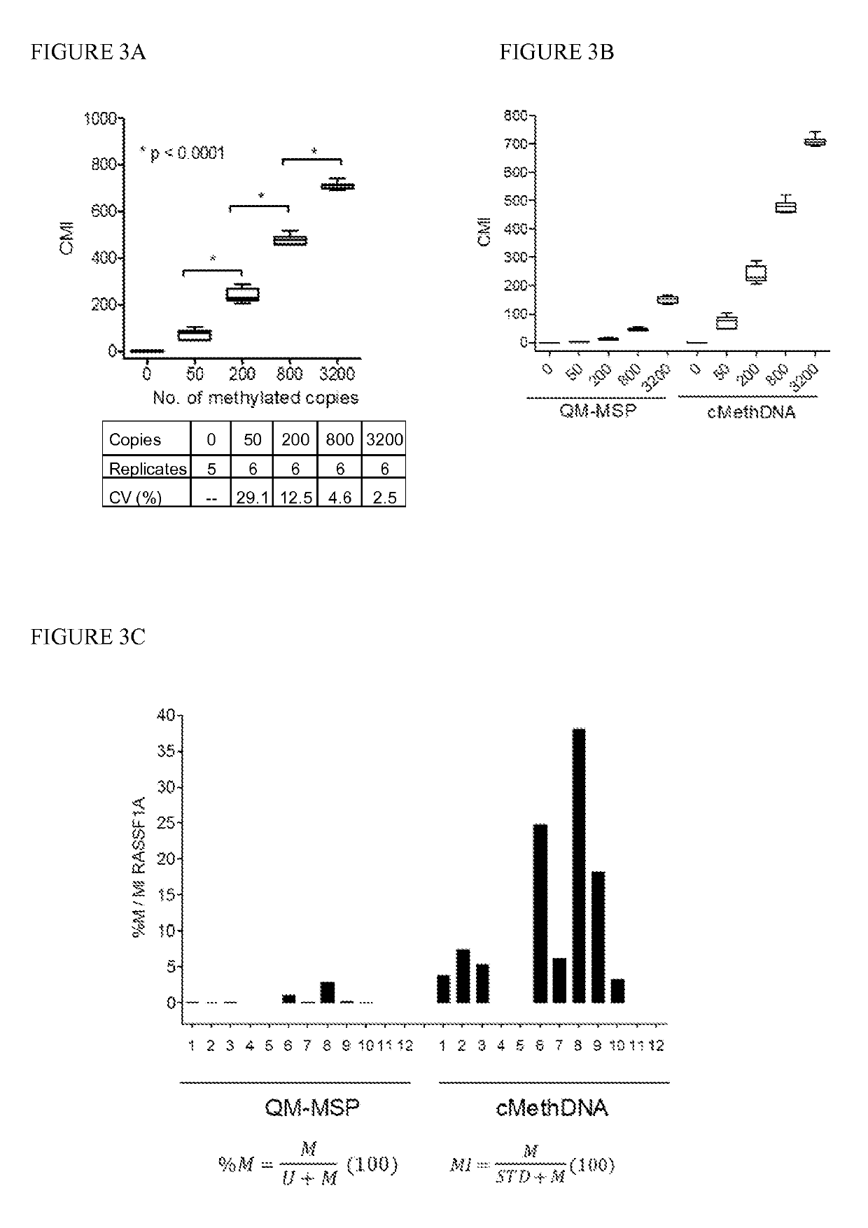
[0092]Technical evaluation of the cMethDNA assay, Analytical linearity and sensitivity. To establish assay linearity and sensitivity, a series of experiments were performed. An optimal procedure was established for isolating minute amounts of circulating DNA by testing three different serum DNA extraction methods (UltraSens Virus Kit, Qiagen; MiniElute Virus Kit, Qiagen, and Quick-gDNA Prep Zymo Research). A master mix of serum from a single normal donor (300 μl serum per assay point) was spiked with a physiological range (0, 50, 200, 800 or 3200 copies) of fully methylated DNA extracted from the MDA-MB-453 breast cancer cell line. The coefficient of variation for each replicate (5-6 replicates) was calculated for each method (FIG. 3A). The lowest level of 50 copies of spiked genomic DNA was detectable by all three methods, and negligible levels of methylated DNA were observed in the normal sera. However, of the three kits tested (FIG. 3A), the MiniElute Virus Spin Kit method displa...
PUM
| Property | Measurement | Unit |
|---|---|---|
| pH | aaaaa | aaaaa |
| temperature | aaaaa | aaaaa |
| volume | aaaaa | aaaaa |
Abstract
Description
Claims
Application Information