Methods and systems for guiding the acquisition of ultrasound images

a technology of ultrasound and acquisition method, applied in the field of guiding the acquisition of ultrasound images, can solve the problems of not having ultrasound training, operator's inability to rely on a consistent location, and others can be more difficult to identify, so as to facilitate the identification of the desired ultrasound scanning path, improve ultrasound scanning, and minimize the distance between the line and the ssc

Active Publication Date: 2009-01-22
ELEKTA AB
View PDF110 Cites 98 Cited by
  • Summary
  • Abstract
  • Description
  • Claims
  • Application Information

AI Technical Summary

Benefits of technology

[0011]Where the SSC and external patient surface contour are contoured on the planning CT, but no planning ultrasound is available, the invention provides may facilitate the identification of the desired ultrasound scanning path using the two contours. In some cases, the desired path is found by tracing a line on the external contour which minimizes the distance between the line and the SSC. The path line may then be projected onto the external surface for visualization, as previously described. Such techniques may improve ultrasound scanning in both the treatment and planning rooms, and is especially useful in the planning room as there is no previous ultrasound image to use as a guide.

Problems solved by technology

One challenge is that radiation therapists, who typically have no ultrasound training, are expected to acquire the ultrasound images during the radiation delivery phase of treatment.
However, there are other potential applications for ultrasound-based IGRT in which an operator cannot rely on a consistent location of the anatomical feature of interest, for example, guiding localized breast-cancer radiotherapy treatments in which the main structure of interest is typically a lumpectomy cavity (i.e. the original site of the surgically-removed tumor).
While some cavities may be easily found using ultrasound, others can be more difficult to identify.
Further, unlike the prostate, which is always in the same general area within the patient, the therapist does not know where to look for the cavity—it can be anywhere within the breast, and therefore requires significant time and experience to find.
This image is typically acquired by either a CT technologist, radiation therapist or radiation oncologist, who also may not have ultrasound experience, giving rise to the same issues as ultrasound-based IGRT.
Because time and trained operators are scarce in a radiotherapy department, finding an anatomical structure such as a lesion cavity using an ultrasound imaging system can be a limiting task in ultrasound-based IGRT of the breast.

Method used


Image

  • Methods and systems for guiding the acquisition of ultrasound images
  • Methods and systems for guiding the acquisition of ultrasound images
  • Methods and systems for guiding the acquisition of ultrasound images

Examples


Embodiment Construction

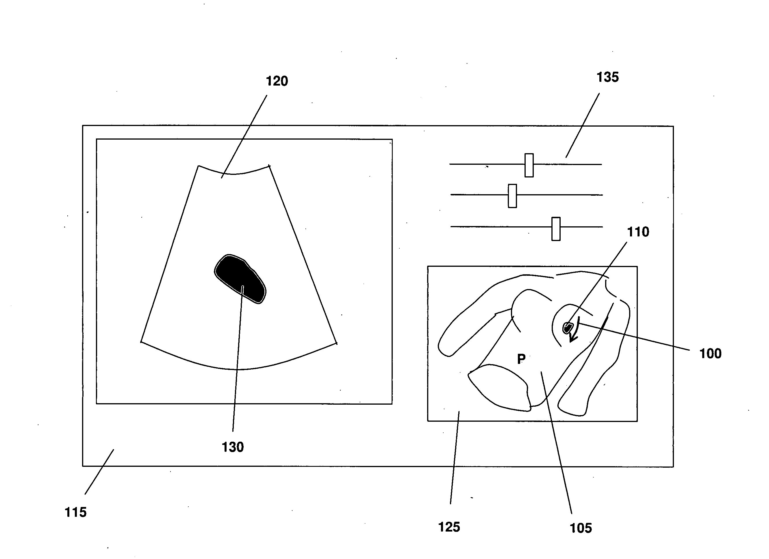
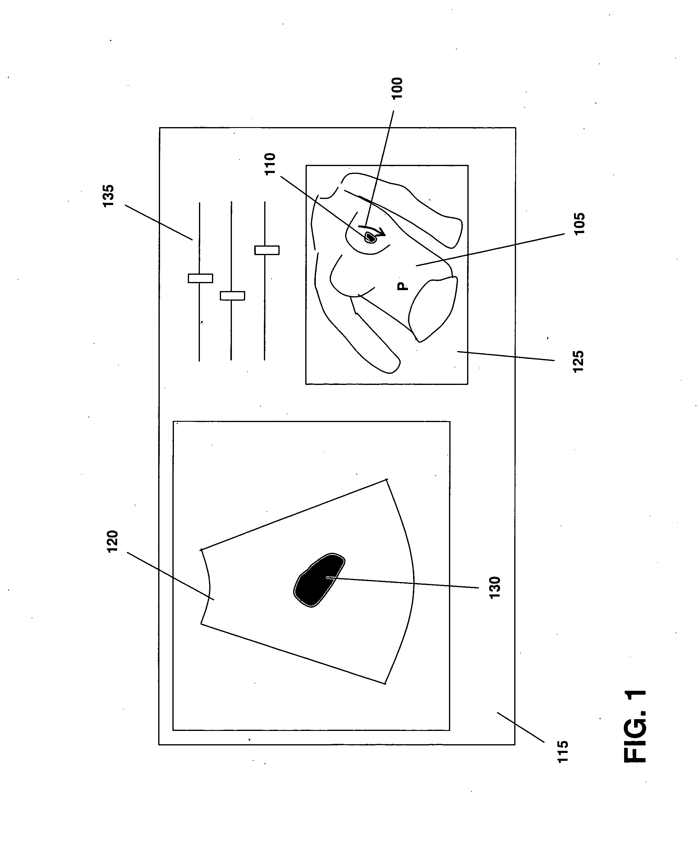
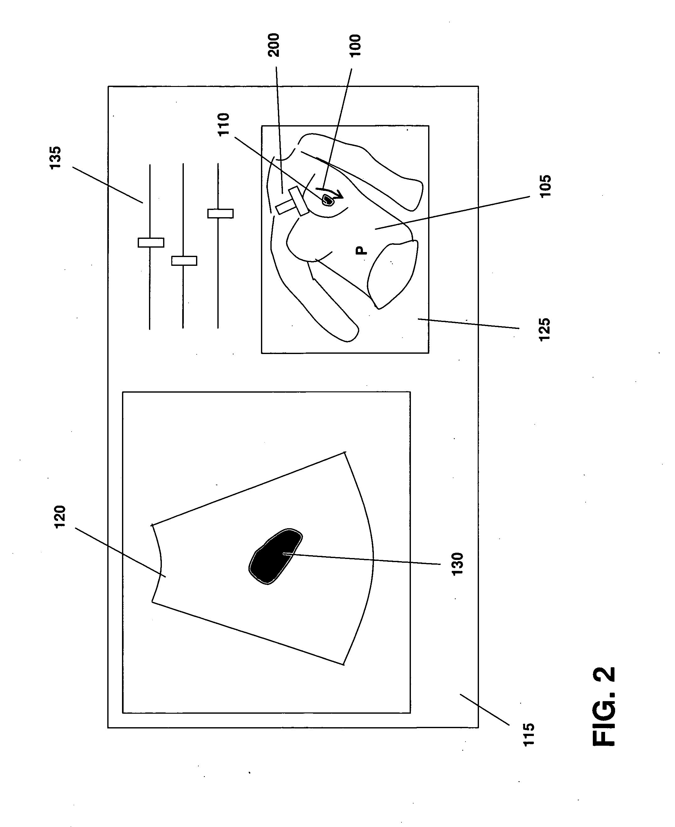
[0019]FIG. 1 illustrates one embodiment of the invention in which a path suggestion line 100 is shown projected onto an external contour 105 of a patient P. A scanning site contour (SSC) 110 is also shown, and may be derived from a three-dimensional surface contour of the anatomical structure of interest, in this case a breast lumpectomy site. In instances in which the SSC lies within the external contour, the contours may be displayed in semi-transparent fashion to allow full visibility of the SSC and other anatomical features. The three-dimensional information may be manipulated to improve viewing (using, for example, conventional visualization devices and / or software), including being be rotated and / or zoomed. The images may also be printed, but preferably are available directly on an ultrasound scanning screen 115 in a treatment planning and / or delivery room. In brief summary, an operator scans the patient using an ultrasound device while reviewing a real-time image 120 to find ...


PUM


Abstract

A method of presenting a suggested path for an ultrasound probe along a patient's surface includes obtaining a three-dimensional, non-ultrasound image of the patient and an ultrasound image of the patient. A treatment area is defined within the within the three-dimensional, non-ultrasound image, and using the images, defining a scanning site contour of an anatomical structure of interest within the patient and an external contour of the patient's surface. A preferred path of the ultrasound scan is projected onto the external contour such that an operator can reproduce the ultrasound scan without knowledge of the anatomical structure of interest.

Description

CROSS-REFERENCE TO RELATED APPLICATIONS[0001]This application claims priority to and the benefit of, and incorporates herein by reference, in its entirety, provisional U.S. patent application Ser. No. 60 / 951,001, filed Jul. 20, 2007.TECHNICAL FIELD[0002]This invention relates to methods for guiding the acquisition of images of anatomical features, and more specifically, guiding the rapid acquisition of ultrasound images that encompass particular features of interest.BACKGROUND INFORMATION[0003]The use of ultrasound imaging systems requires advanced training and is most often performed by sonographers or physicians who have an intimate knowledge of the anatomy under study, as well as the expected appearance of anatomical features in an ultrasound image. For example, in order to pinpoint the correct probe positioning to acquire an image of a desired anatomical structure (e.g., an organ, lesion or tumor), an operator must coordinate moving the ultrasound probe to the correct location o...

Claims


Application Information

Patent Timeline
Patent Type & Authority Applications(United States)
IPC IPC(8): A61B8/13
CPCA61B8/0825A61B8/14A61B8/4416A61B8/4245A61B8/4218
Inventor LACHAINE, MARTINFALCO, TONY
Owner ELEKTA AB
Why Eureka
Try Eureka