Medical Imaging Machine and Methods of Use

a technology of medical imaging machine and method, applied in the field of medical imaging, can solve problems such as safety issues and availability problems

Inactive Publication Date: 2012-06-14
HELIOCENTRIC TECH
View PDF0 Cites 10 Cited by
  • Summary
  • Abstract
  • Description
  • Claims
  • Application Information

AI Technical Summary

Benefits of technology

[0010]The present inventor realized in an inventive moment that if, during the imaging procedure, stable isotopes that might respond to a focused external stimulation could be employed instead of single-state active radioisotopes, better control over the imaging process and improvement in safety might result. The inventor therefore constructed a unique medical imaging system for clinical use that allowed non-radioactive (stable) tracer isotopes to be used as tracer elements in place of characteristic radioactive tracer isotopes, the stable isotopes activated in a controlled manner using an external neutron generator fuel. A significant improvement system process control and material handling efficiency results, with no impediment to image quality or imaging time created.
[0015]According to another aspect of the present invention, In a system for medical imaging, the system including a neutron generator, a gantry for containing an imaging subject containing at least one non-radioactive tracer isotope, and at least one gamma ray camera electrically connected to a processor-based data acquisition system, a method for processing a gamma ray spectrum or image to form a deconvolved two-dimensional or three-dimensional image including the steps (a) aided by software running on the data acquisition system, applying a neutron shelf-shielding correction factor to the gamma ray spectrum or image, (b) aided by the software of step (a), applying a gamma ray attenuation correction factor to the gamma ray spectrum or image, (c) filtering the image data of noise, and (d) processing the image data of step (c) to improve discernability of the image.

Problems solved by technology

The problem stated above is that patient and operator safety along with adequate availability of tracer materials are desirable characteristics for a nuclear medical imaging system, but many of the conventional means for obtaining medical images in clinical settings, such as single photon emission computed tomography (SPECT) imaging systems, also create safety issues and availability issues relative to the unpredictable availability of tracer isotopes for medical industry consumption.

Method used


Image

  • Medical Imaging Machine and Methods of Use
  • Medical Imaging Machine and Methods of Use
  • Medical Imaging Machine and Methods of Use

Examples


Embodiment Construction

[0025]The inventors provide a unique system and accompanying methods for medical imaging that enable construction of deconvolved medical images for clinical use. The present invention will be described in enabling detail using the following examples, which may describe more than one relevant embodiment falling within the scope of the present invention.

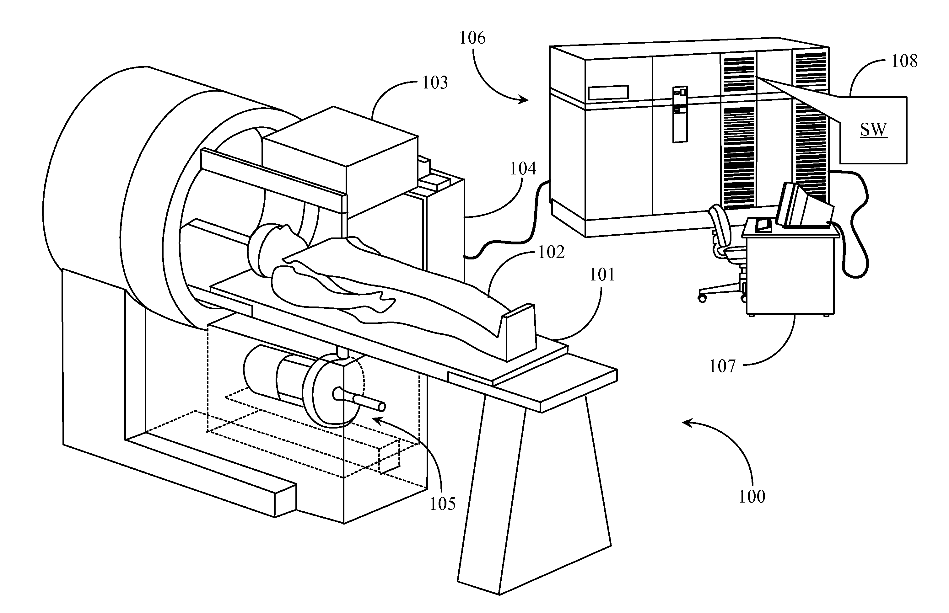
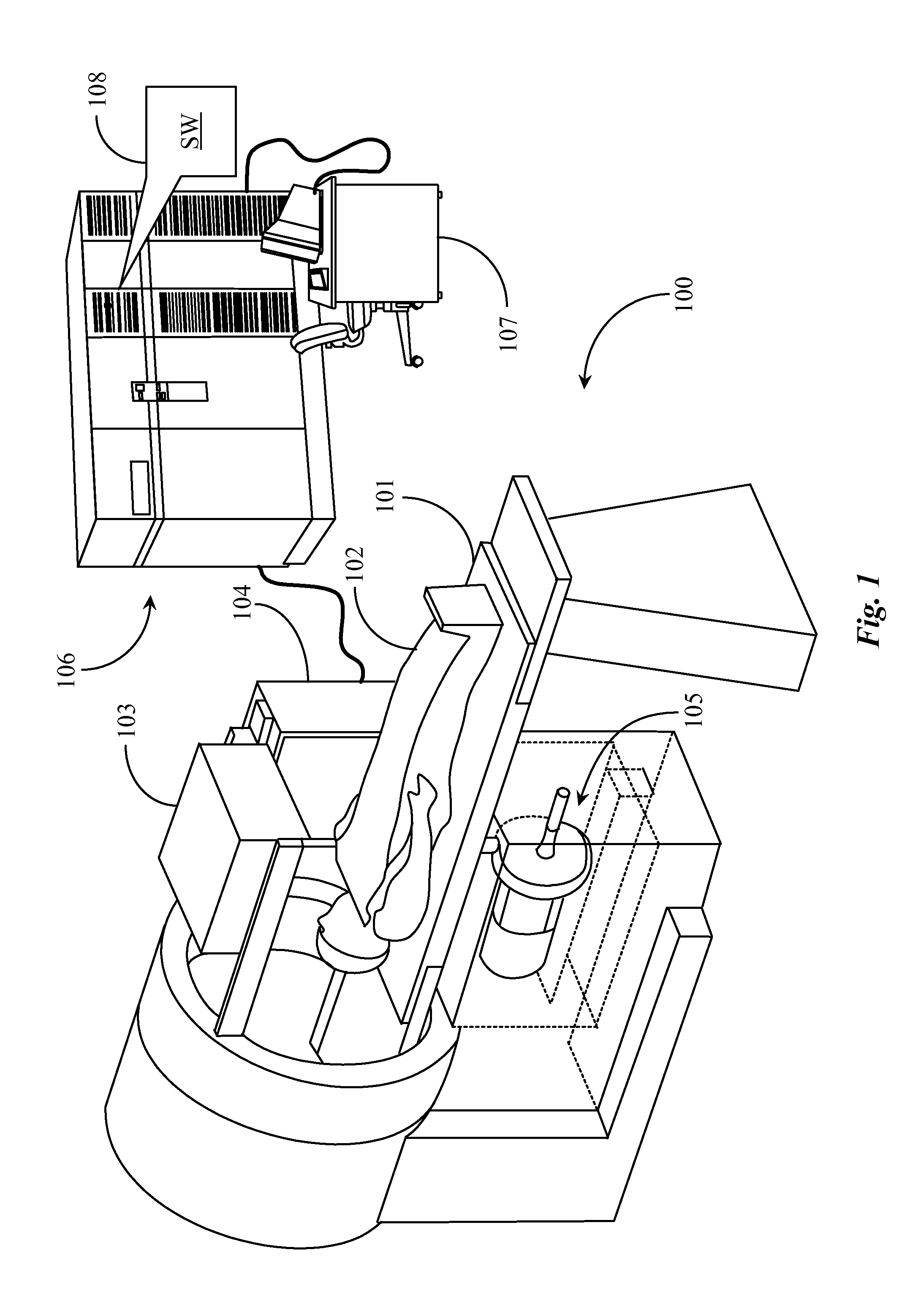
[0026]FIG. 1 is a perspective view of a neutron-activated single photon emission computed tomography (nSPECT) system 100. The nSPECT system 100 is a hybrid medical imaging system that uses prompt gamma-ray neutron activation (PGNA) to activate non-radioactive tracer isotopes rather than typical gamma ray detection associated with injected radioactive tracer isotopes typically used in SPECT imaging.

[0027]A gantry 101 similar to that of a SPECT imaging system is illustrated in perspective and supports an imaging subject 102 on the gantry table for medical imaging. The rear portion of gantry 101 includes at least one rotable ring for rota...


PUM


Abstract

A system for medical imaging is provided and includes at least one neutron generator having a neutron generator fuel material and at least one neutron moderator material, a gantry for stationing an imaging subject, a neutron collimator attached to the neutron generator, the collimator disposed between the neutron moderator and the imaging subject, at least one gamma ray camera electrically connected to a processor-based data acquisition system, and software executing on the processor-based data acquisition system from a non-transitory physical medium, the software providing a first function for producing at least one gamma ray spectrum or image, a second function for applying correction factors to the gamma ray spectra or images, and a third function for analyzing the corrected gamma ray spectra or images to process one or more clinically relevant images of one or more targeted or general areas of the imaging subject.

Description

CROSS-REFERENCE TO RELATED APPLICATIONS[0001]The present invention claims priority to a U.S. provisional patent application Ser. No. 61 / 422,869, entitled “Neutron Activated Single Photon Emission Computed Tomography”, filed on Dec. 14, 2010, disclosure of which is incorporated in this specification at least by reference.BACKGROUND OF THE INVENTION[0002]1. Field of the Invention[0003]The present invention is in the field of medical imaging, particularly nuclear imaging systems and pertains particularly to methods and apparatus for obtaining medical image data using non-radioactive tracer isotopes controllably activated by external neutron emission.[0004]2. Discussion of the State of the Art[0005]The field of nuclear medicine includes nuclear imaging systems such as single photon emission computed topography (SPECT) systems. In SPECT imaging, radioactive isotopes serving as tracer elements are injected into the subject's bloodstream and are later tracked through detection of the gamma...

Claims


Application Information

Patent Timeline
Patent Type & Authority Applications(United States)
IPC IPC(8): G06K9/00
CPCG01T1/1642A61B6/4258A61B6/037
Inventor MCRAE, GLENNFABER, DANIELBELL, ADAM
Owner HELIOCENTRIC TECH
Why Eureka
Try Eureka