Method for providing a 3D image data record of a physiological object with a metal object therein

Active Publication Date: 2012-09-06
SIEMENS HEALTHCARE GMBH
View PDF4 Cites 1 Cited by
  • Summary
  • Abstract
  • Description
  • Claims
  • Application Information

AI Technical Summary

Benefits of technology

[0006]It is the object of the present invention to propose a method for providing a 3D image data record for the purpose of recording an image of a physiologi

Problems solved by technology

Since only one representation of the needle from one single perspective was previously available, at best two with

Method used


Image

  • Method for providing a 3D image data record of a physiological object with a metal object therein
  • Method for providing a 3D image data record of a physiological object with a metal object therein

Examples


Embodiment Construction

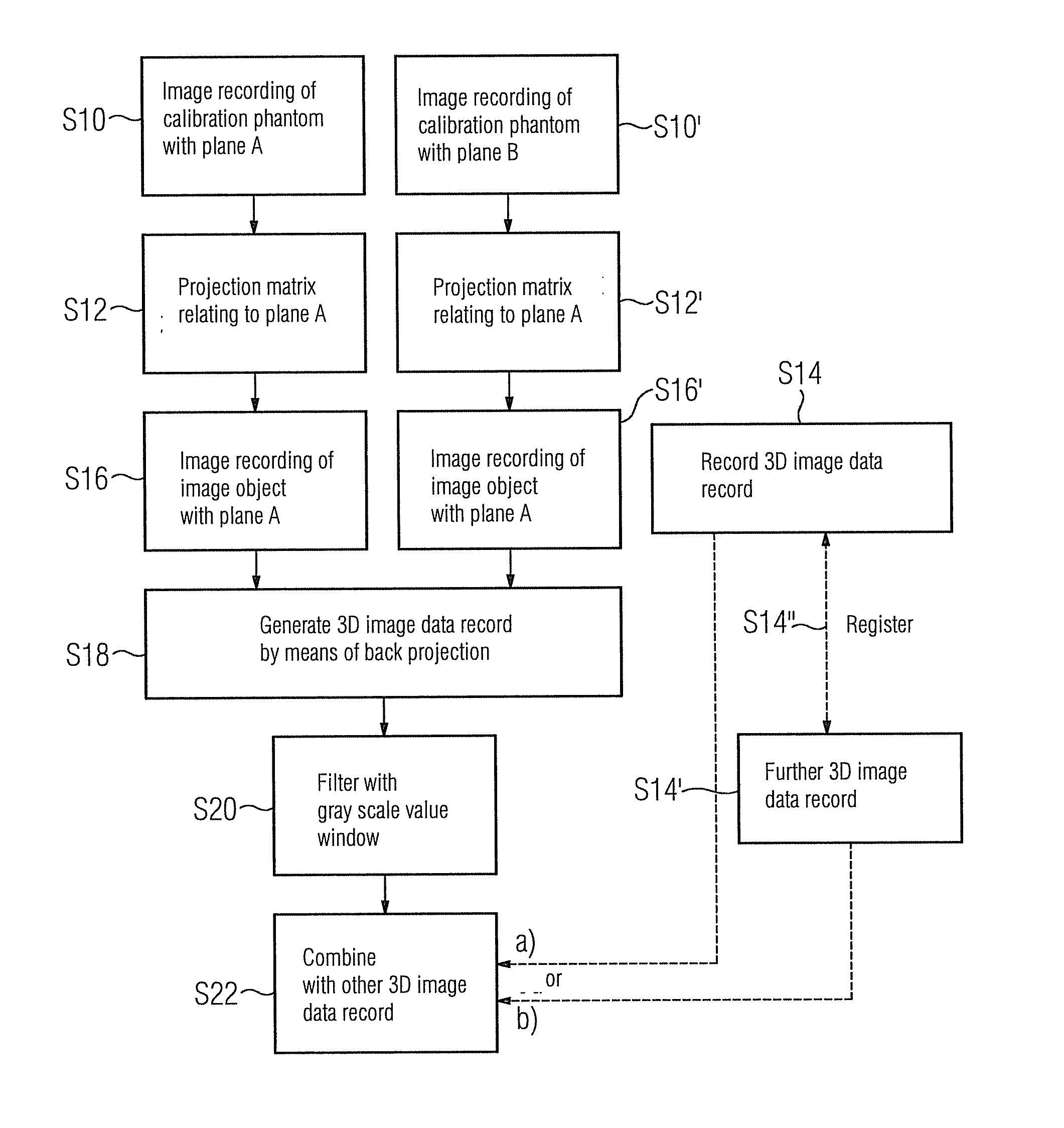
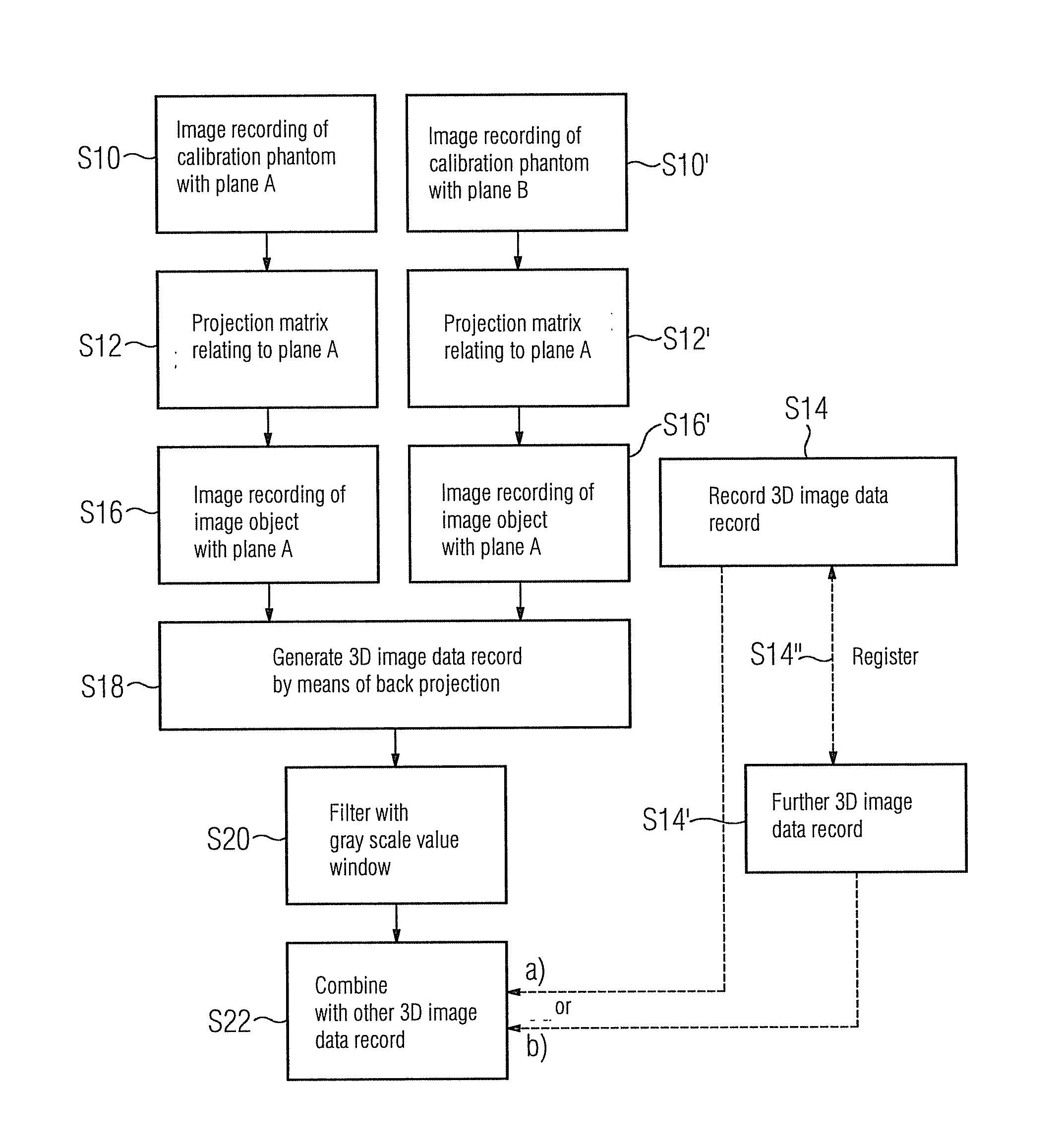
[0020]It is currently assumed that a biplane x-ray system exists, i.e. an x-ray image recording apparatus having two x-ray image recording units, each of which includes an x-ray source and an x-ray detector. The tem, “biplane system” means that the one x-ray detector can lie in a plane and the other x-ray detector can lie in another plane at the same time. The one plane is currently referred to as “plane A”, the other as “plane B”, whereby these planes are preferably to stand precisely at an angle of 90° relative to one another.

[0021]A calibration phantom is now initially brought into the biplane x-ray system and an image is recorded, namely in steps S10 and S10′ which are to be completed synchronously by means of the two x-ray image recording units. The respective projection matrix relating to the plane and thus to the position of the x-ray detector and also the x-ray source can be calculated in a manner known per se in step S12 and also in step S12′ with the aid of the thus obtain...


PUM


Abstract

A method for providing a 3D image data record of a physiological object with a metal object therein is proposed. To enable an image of the metal object in the physiological object, for instance a biopsy needle in a human patient to be recorded as a 3D image in this patient, two 2D x-ray images of the patient are obtained with the needle with the aid of a biplane x-ray system, and back projection allows a 3D image data record to be generated, which is then subject to a filtering with a gray-scale value window. After filtering, information relating to the position and shape of the metal object in space is obtained. The thus obtained first 3D image data record can be combined with another 3D image data record, in particular with a 3D image data record of the patient without the metal object obtained with the same biplane x-ray system.

Description

CROSS REFERENCE TO RELATED APPLICATIONS[0001]This application claims priority of German application No. 10 2011 005 119.8 filed Mar. 4, 2011, which is incorporated by reference herein in its entirety.FIELD OF INVENTION[0002]The invention relates to a method for providing a 3D image data record for the purpose of recording an image of a physiological object with a metal object located therein. The physiological object may in particular be a patient and the metal object may be an object or part of an object, which is inserted into the patient within the scope of a medical treatment. The metal object may therefore include a needle inserted into the body of the patient, or also a guidewire.BACKGROUND OF INVENTION[0003]Thus for instance, during the biopsy of a tumor, a hollow needle is pushed into the patient in order to take tissue samples of the tumor which can then be analyzed later in a lab. It must be ensured here that the removed sample is part of the non-necrotic tumor mass. The n...

Claims


Application Information

Patent Timeline
IPC IPC(8): G06K9/00
CPCA61B6/02A61B6/4014A61B6/583A61B2019/5295A61B19/52A61B2019/5238A61B10/0233A61B6/022A61B90/36A61B2090/367A61B2090/376
Inventor BENDER, FREDERIKROYALTY, KEVIN
Owner SIEMENS HEALTHCARE GMBH
Why Eureka
Try Eureka