Removal of embedding media from biological samples and cell conditioning on automated staining instruments

Inactive Publication Date: 2003-04-08
VENTANA MEDICAL SYST INC
View PDF28 Cites 285 Cited by
  • Summary
  • Abstract
  • Description
  • Claims
  • Application Information

AI Technical Summary

Benefits of technology

The present invention relates to solutions, methods and instruments for cell or tissue conditioning, which improves the accessibility of molecules in bi

Problems solved by technology
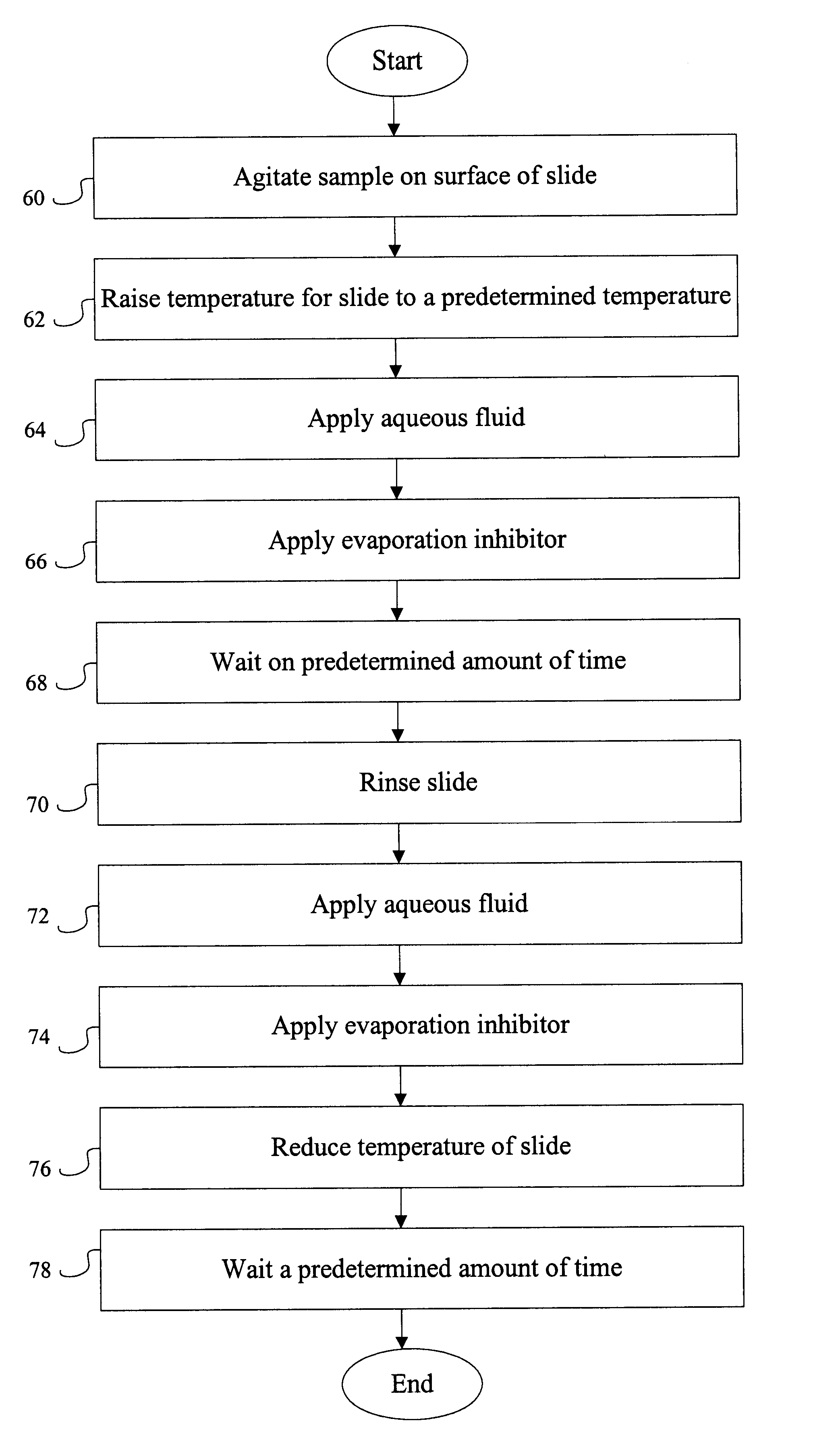
Without preservation, tissue samples rapidly deteriorate such that their use in diagnostics is compromised shortly after removal from their host.
Some antigens have particular pH requirements such that adequate results will only be achieved in a narrow pH range.
Although the chemical components of the antigen retrieval solution, including metal ions, may play a role as possible co-factors in the microwave heating procedure, thus far, no single chemical has been identified that is both essential and best for antigen retrieval.
These organic solvents are very volatile causing a variety of problems includ

Method used


Image

  • Removal of embedding media from biological samples and cell conditioning on automated staining instruments
  • Removal of embedding media from biological samples and cell conditioning on automated staining instruments
  • Removal of embedding media from biological samples and cell conditioning on automated staining instruments

Examples


Example

Example 1

Automated "Exposing" and "Cell Conditioning"with Biological Samples Stained with H&E

Biological samples, including breast, stomach, brain, tonsil and kidney, that had been embedded in paraffin were exposed according to the following procedure: slides containing the above referenced biological sample were placed on an automated instrument (Ventana Medical Systems, Inc., Tucson, Ariz.) and subjected to the exposing protocol described below. Generally, the slides containing paraffin embedded biological samples were dry heated to 65.degree. C. for six (6) minutes then rinsed with any of the following: 1) 1.times. citrate buffer, 2) de-ionized water, 3) 10 mM phosphate buffer (pH=6.3), or 4) 10 mM Tris-HCl buffer (pH=7.4) each containing 0.1% Triton X-100.

EXPOSING PROTOCOL 1 1. Incubate for 2 minutes 2. Rinse slide 3. Adjust slide volume and apply LIQUID COVERSLIP.TM. 4. Incubate for 6 minutes 5. Rinse slide 6. Adjust slide volume and apply LCS.TM. 7. Increase temperature to 65.0...

Example

Example 2

Automated "Exposing" of Biological Samples with Simultaneous "Cell Conditioning"

Biological samples of kidney and tonsil that had been formalin fixed and embedded in paraffin were exposed according to the protocol described in Example 1. After automated exposing, the biological sample was subjected to the automated DAB paraffin protocol used for immunohistochemical staining. The protocol for DAB staining is described below:

DAB Protocol 1. Incubate for 2 minutes 2. Rinse slide 3. Adjust slide volume and apply LCS.TM. 4. Rinse slide 5. Adjust slide volume and apply LCS.TM. 6. Rinse slide 7. Adjust slide volume and apply LCS.TM. 8. Apply one drop of inhibitor 9. Incubate for 4 minutes 10. Adjust slide volume and apply LCS.TM. 11. Apply one drop of primary antibody 12. Incubate for 32 minutes 13. Adjust slide volume and apply LCS.TM. 14. Apply one drop of Biotinylated Ig 15. Incubate for 8 minutes 16. Rinse slide 17. Adjust slide volume and apply LCS.TM. 18. Apply one drop of A ...

Example

Example 3

Two Step Automated "Exposing" and "Cell Conditioning"

Biological samples of tonsil and breast that had been preserved in paraffin and treated with formaldehyde were treated by the following protocol:

Exposing and Cell Conditioning Protocol 5 1. Incubate for 2 minutes 2. Increase thermofoil temperature to 65.0.degree. C. 3. Incubate for 6 minutes 4. Rinse slide and apply LCS.TM. 5. Incubate for 6 minutes 6. Rinse slide and apply LCS.TM. 7. Increase thermofoil temperature to 100.0.degree. C. 8. Adjust slide volume and apply LCS.TM. 9. Rinse slide 10. Adjust slide volume and apply LCS.TM. 11. Incubate for 4 minutes 12. Adjust slide volume and apply LCS.TM. 13. Incubate for 4 minutes 14. Adjust slide volume and apply LCS.TM. 15. Incubate for 4 minutes 16. Adjust slide volume and apply LCS.TM. 17. Incubate for 4 minutes 18. Adjust slide volume and apply LCS.TM. 19. Incubate for 4 minutes 20. Adjust slide volume and apply LCS.TM. 21. Incubate for 4 minutes 22. Adjust slide volume a...


PUM


Abstract

The present invention provides reagents for use in an automated environment for cell conditioning of biological samples wherein the cells or tissues are predisposed for access by reagent molecules for histochemical and cytochemical staining procedures. The reagents include components optimized to facilitate molecular access to cells and cell constituents within the biological sample. The present invention also provides reagents for use in an automated environment for removing or etching embedding media by exposing a biological sample to be stained in histochemical or cytochemical procedures without the dependence on organic solvents. The reagents include components optimized to facilitate removal or etching of the embedding media from the biological sample.

Description

BACKGROUND OF THE INVENTION1. Field of the InventionThe present invention relates to solutions methods, and instruments for conditioning cells or tissues so as to increase the accessibility of various molecules to their respective targets and generally to improve tissue and cell readability of biological samples on automated instruments prior to immunohistochemical (IHC), in situ hybridization (ISH) or other histochemical or cytochemical manipulations.2. Summary of the Related ArtAll patents, patent applications and non-patent articles or references mentioned herein are hereby incorporated by reference to the extent that they are not contradictory.The diagnosis of disease based on the interpretation of tissue or cell samples taken from a diseased organism has expanded dramatically over the past few years. In addition to traditional histological staining techniques and immunohistochemical assays, in situ techniques such as in situ hybridization and in situ polymerase chain reaction a...

Claims


Application Information

Patent Timeline
IPC IPC(8): G01N1/36G01N33/48G01N1/28
CPCG01N1/36Y10T436/107497Y10T436/25125Y10T436/25375
Inventor CHRISTENSEN, KIMBERLYMACREA, ETHEL R.SEBASTIAO, NOEMIWARD, GLEN
Owner VENTANA MEDICAL SYST INC
Why Eureka
Try Eureka