Device and method for endoluminal therapy

a technology of endoluminal plasty and device, which is applied in the field of endoluminal plasty, can solve the problems of difficult endoluminal plasty, difficult endoluminal plasty, and high variability in the thickness of the mucosal layer, and achieve the effect of minimizing the dilation of the pouch or restrictive outlet over tim

Inactive Publication Date: 2010-02-23
KELLEHER BRIAN +1
View PDF10 Cites 606 Cited by
  • Summary
  • Abstract
  • Description
  • Claims
  • Application Information

AI Technical Summary

Benefits of technology

[0015]The preferred methods and devices described herein provide for advanced endoluminal procedures involving hollow organs. Such advanced procedures involve selective engagement of a target layer within the wall of a hollow organ, approximation of multiple regions of the wall, and safe and reliable placement of securement elements through the full thickness of the wall without endangering structures surrounding the hollow organ. Specific methods and devices described enable endoluminal procedures such as gastroplasty for the treatment of obesity and GERD, involving the formation of a gastric pouch or partition and a restrictive outlet. Further methods and devices provide for minimizing the dilation of the pouch or restrictive outlet over time.
[0018]In yet another group of preferred embodiments of the invention, methods and devices are described for approximating two or more regions of a wall of a hollow organ. In at least one preferred embodiment for a stomach organ, the muscularis wall layer is selectively engaged in two or more regions using the methods and devices described previously, and then the regions are pulled together. In another preferred embodiment, the walls are pulled into the lumen to create a safety gap to allow safe and reliable deployment of securement elements through the wall where the securement elements are then pulled together to approximate the regions. In another embodiment, the walls are approximated and then pulled into the lumen to create a safety gap prior to deployment of a securement element through both regions of the wall.
[0022]In another group of preferred embodiments, methods are disclosed for treating the walls of the stomach in the vicinity of a gastric pouch and restrictive outlet to minimize the amount of dilation that may occur. Such methods include modifying the properties of the stomach wall so as to create scarification or shrinkage of the tissue layers.

Problems solved by technology

Until now, there have been no practical devices available to enable procedures such as endoluminal plasty in a safe and reliable manner.
The most challenging part of such a procedure is the placement of durable securement elements, such as sutures, through the full thickness of the organ walls in order to secure portions of the organ together or to attach elements to the wall.
The reason securement element placement is challenging is that the organ wall may consist of multiple layers, each having unique physical properties.
The mucosa typically comprises a plurality of folds, called rugal folds, or rugae, which make the thickness of the mucosal layer highly variable and unpredictable.
This presents a challenge for endoluminal therapy, however, since the combined thickness of the three layers of the stomach wall is normally unpredictable.
When the desired layer of a wall is the outermost layer, surrounding structures may be in danger of being engaged by such securement element or deployment device.
Another challenge of endoluminal devices intended for use in organ therapy is the engagement and movement of regions of the organ wall in order to approximate and secure multiple locations.
It may require a significant amount of force to move a region of the organ wall, pulling by the innermost layer may tear it.
Further, the process of approximating two regions of the wall may require multiple instruments.

Method used


Image

  • Device and method for endoluminal therapy
  • Device and method for endoluminal therapy
  • Device and method for endoluminal therapy

Examples


Embodiment Construction

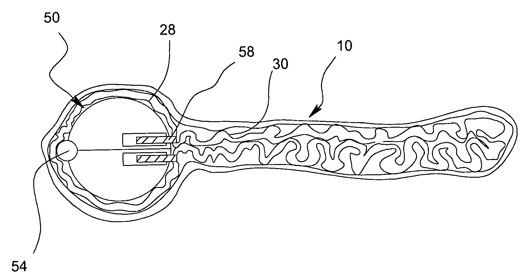
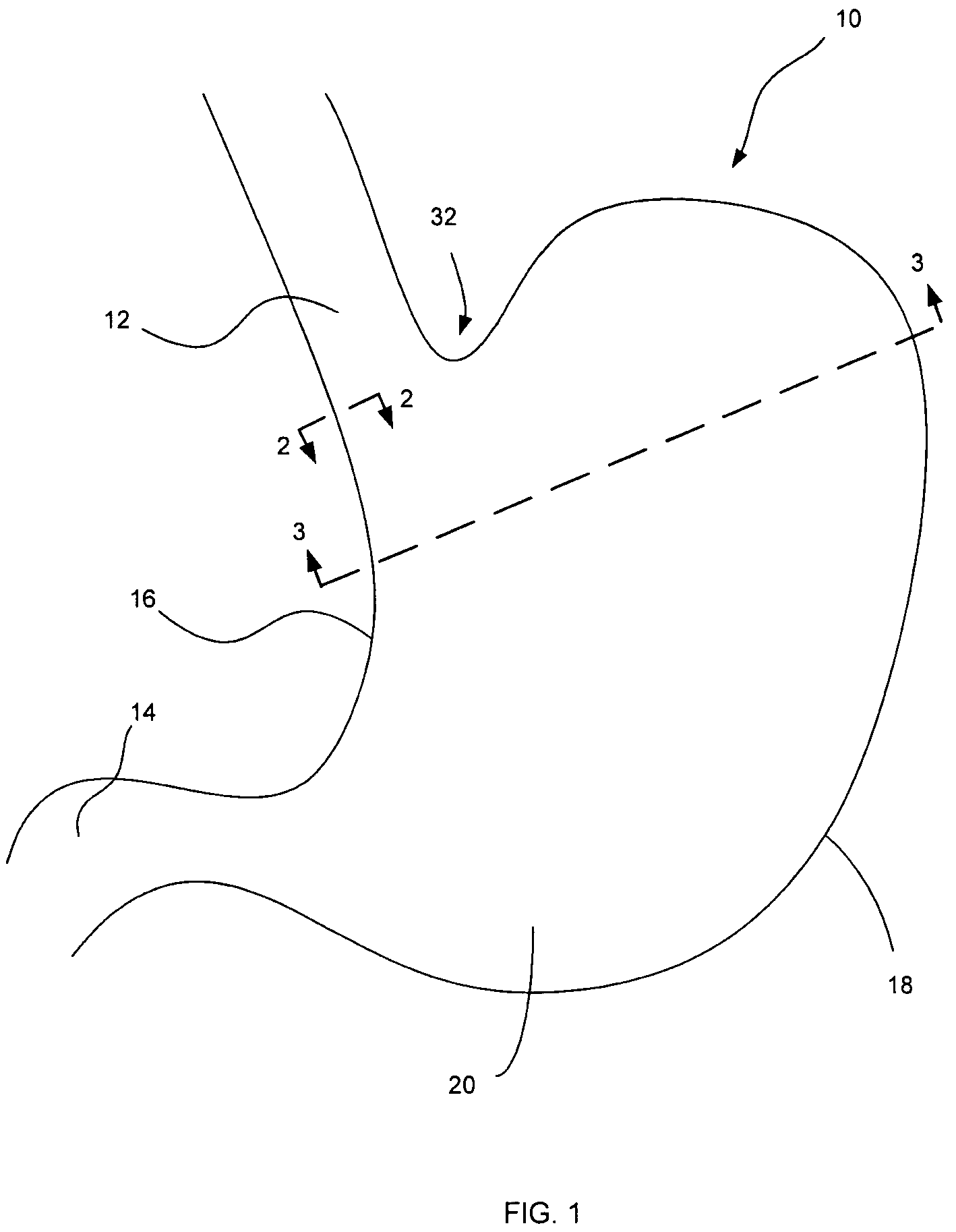
[0092]The present invention relates to endoluminal therapy and particularly to methods and devices for manipulation of the walls of luminal organs such as the stomach. Before describing embodiments of the devices and methods of the invention, an exemplary luminal organ, a stomach 10 will first be described in more detail. It is understood that the method and devices of the present invention may be applied to any luminal organ. Application of the present invention a stomach 10 is thus for illustrative purposes only. FIGS. 1, 2 and 3 illustrate a representation of the normal anatomy of a stomach 10.

[0093]As shown if FIG. 1, the exemplary luminal organ, the stomach 10 includes an upper opening at the esophagus 12, an Angle of His 32 at the junction between esophagus 12 and stomach 10, and a lower opening at the pylorus 14 which communicates with the small intestine (not shown). For reference, the stomach 10 further includes a lesser curvature 16 and a greater curvature 18 as well as po...


PUM


Abstract

A device and method for selectively engaging or penetrating a layer of a luminal organ wall where the luminal organ wall has a plurality of layers including an outermost layer and an innermost layer adjacent to the lumen of the organ. The device and method select one of the plurality of layers of the organ wall other than the innermost layer and deploy from within the lumen of the organ a tissue device through the innermost layer to a specific depth to engage or penetrate the selected one of the plurality of layers. The device and method may be employed to create luminal pouches or restrictive outlets. In a stomach organ, the device and methods may be employed to treat obesity by forming a gastric pouch with or without a restrictive outlet.

Description

CROSS REFERENCE TO RELATED APPLICATIONS[0001]This application is related to Provisional Patent Application 60 / 409,838, filed Sep. 9, 2002, which is hereby incorporated by reference for its teachings.BACKGROUND[0002]1. Field of the Invention[0003]The invention relates to endoluminal therapy, and, in particular, to methods and devices for securement of hollow organs, including gastric restriction procedures in the stomach for treating obesity and gastroesophageal reflux disease (GERD).[0004]2. Description of the Related Art[0005]Flexible endoscopic therapy of the hollow organs such as the stomach, intestines and esophagus has been shown to have fewer complications and faster recovery times than traditional or laparoscopic surgery. As such, there is a growing interest in expanding the capabilities of such therapy. In particular, there is a desire to perform organ modification, for example gastroplasty, from within the lumen of the organ through a trans-luminal route using flexible endo...

Claims


Application Information

Patent Timeline
Patent Type & Authority Patents(United States)
IPC IPC(8): A61B17/04A61B17/02A61B17/06A61B17/064A61B17/068A61B17/072A61B17/08A61B17/115A61B17/28A61F5/00
CPCA61B17/0218A61B17/0401A61B17/0469A61B17/0482A61B17/0491A61B17/068A61B17/072A61F5/0086A61B17/0206A61B17/0487A61B17/0644A61B17/1152A61B2017/00827A61B2017/0409A61B2017/0417A61B2017/0458A61B2017/047A61B2017/0472A61B2017/0496A61B2017/06052A61B2017/07214A61B2017/2905A61B2017/2943
Inventor KELLEHER, BRIANSTONE, CORBETT
Owner KELLEHER BRIAN
Why Eureka
Try Eureka