System, apparatus and methods for providing a single use imaging device for sterile environments

a technology of sterile environment and imaging device, applied in the field of imaging device, can solve the problems of unsafe use and expired imaging devi

Active Publication Date: 2014-02-11
DEPUY SYNTHES PROD INC
View PDF200 Cites 22 Cited by
  • Summary
  • Abstract
  • Description
  • Claims
  • Application Information

AI Technical Summary

Benefits of technology

[0012]In an embodiment of an active imaging device may be attached to a control unit. The control unit will check the last sterilization date and ensure that the imaging device is no older than a predetermined safety date. If the imaging device is older than the required date, an onscreen warning will tell the user that the imaging device has expired and is unsafe for use. These features will protect the patient and the doctor from using a non-sterile imaging device.

Problems solved by technology

The imaging device may display an onscreen message telling the user that the camera has already been used and will not allow current operation.
If the imaging device is older than the required date, an onscreen warning will tell the user that the imaging device has expired and is unsafe for use.

Method used


Image

  • System, apparatus and methods for providing a single use imaging device for sterile environments
  • System, apparatus and methods for providing a single use imaging device for sterile environments
  • System, apparatus and methods for providing a single use imaging device for sterile environments

Examples


Embodiment Construction

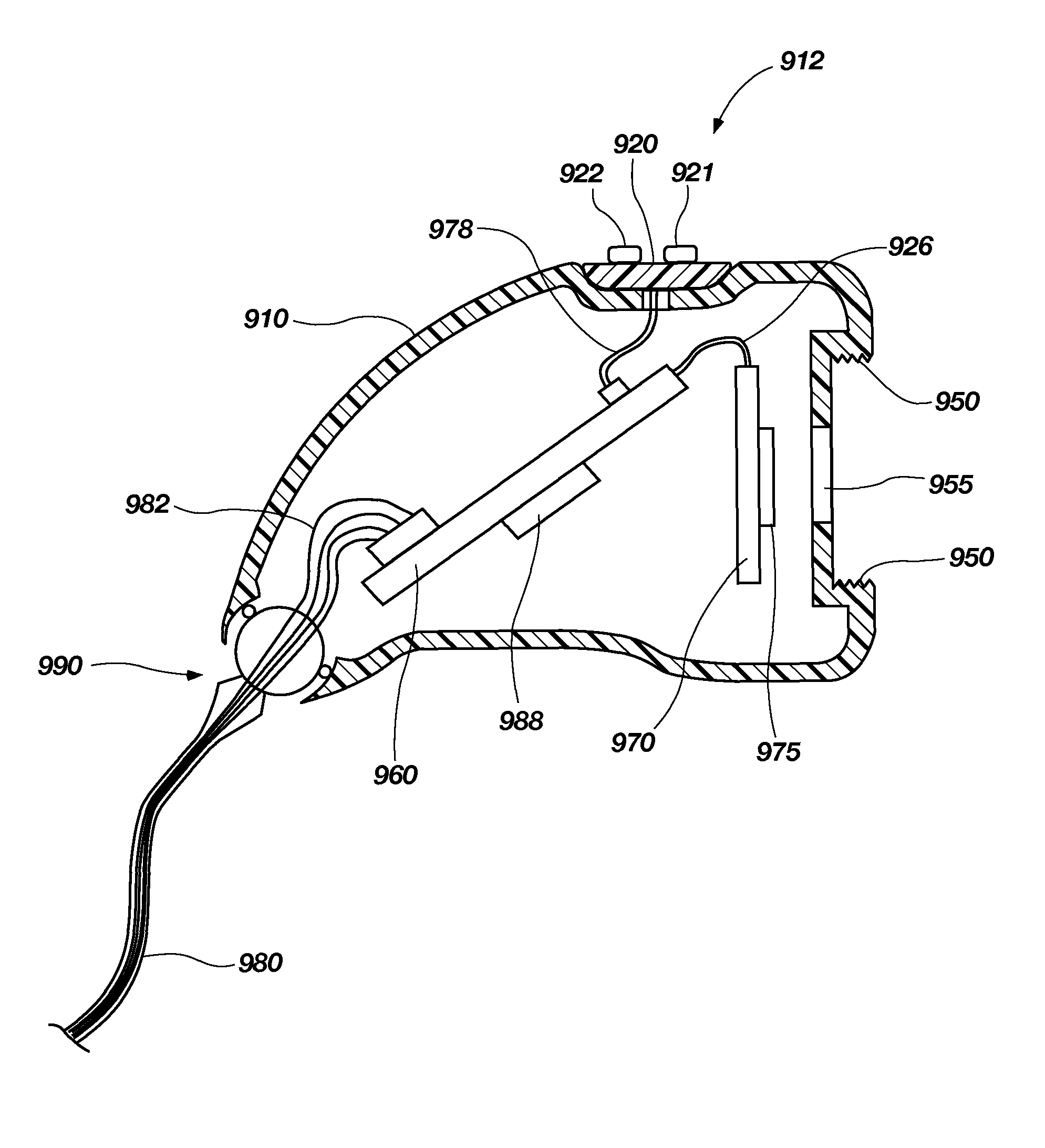
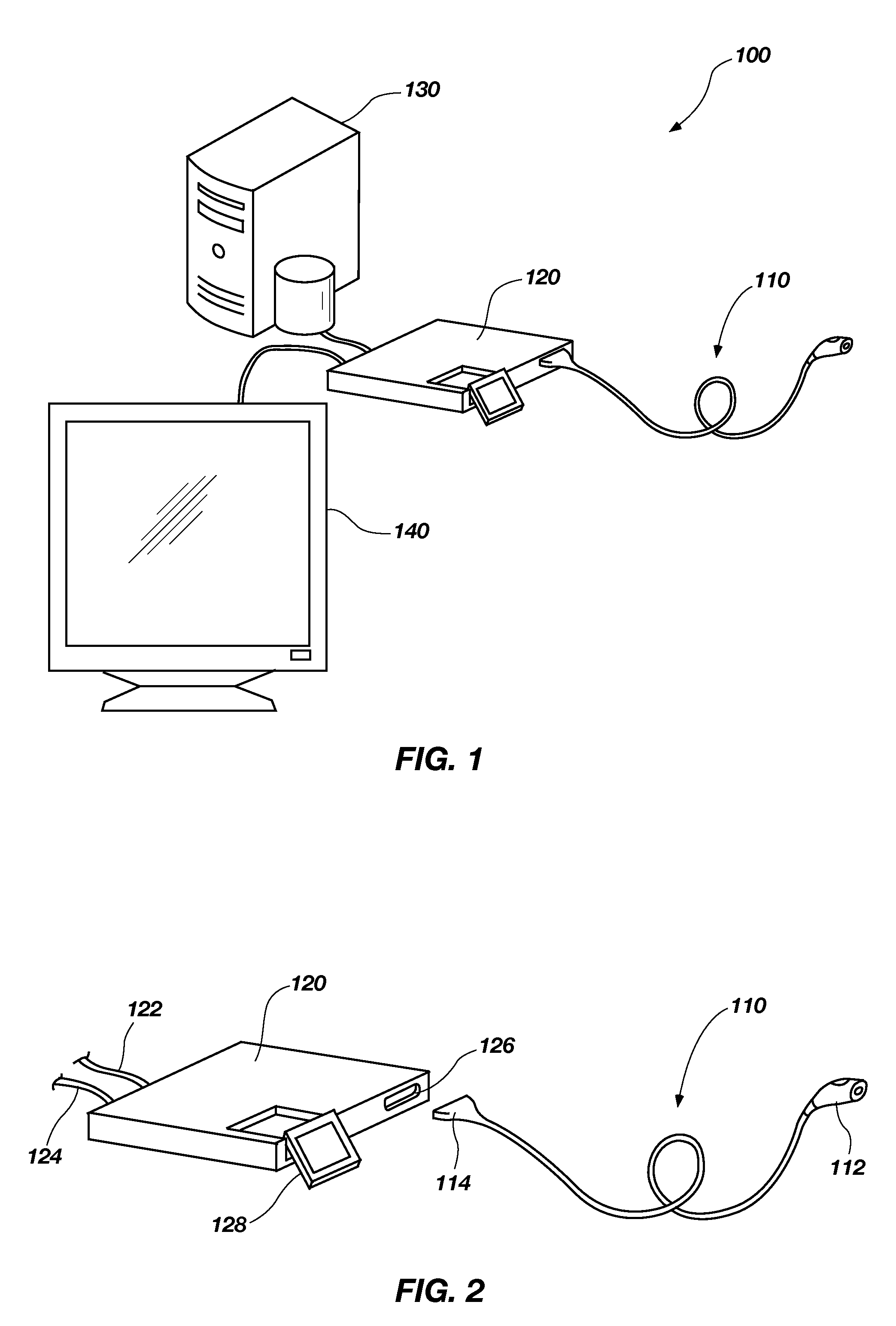
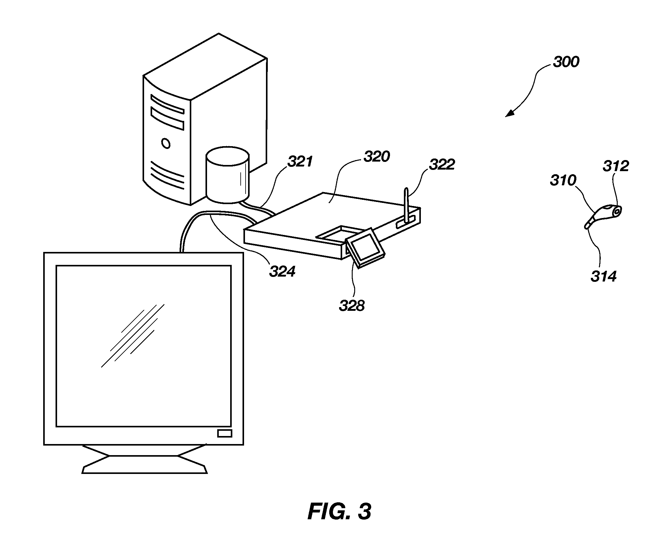
[0037]For the purposes of promoting an understanding of the principles in accordance with the disclosure, reference will now be made to the embodiments illustrated in the drawings and specific language will be used to describe the same. It will nevertheless be understood that no limitation of the scope of the disclosure is thereby intended. Any alterations and further modifications of the inventive features illustrated herein, and any additional applications of the principles of the disclosure as illustrated herein, which would normally occur to one skilled in the relevant art and having possession of this disclosure, are to be considered within the scope of the disclosure claimed.

[0038]Before the devices, systems, methods and processes for providing and reclaiming single use imaging devices are disclosed and described, it is to be understood that this disclosure is not limited to the particular embodiments, configurations, or process steps disclosed herein as such embodiments, conf...


PUM


Abstract

A system, apparatus and methods for providing a single use imaging device for sterile environments is disclosed and described. A single use high definition camera used for general purpose surgical procedures including, but not limited to: arthroscopic, laparoscopic, gynecologic, and urologic procedures, may comprise an imaging device that is a sterile and designed to ensure single use. The imaging device may have a single imaging sensor, either CCD (charge coupled device) or CMOS (complementary metal oxide semiconductor), encased in a housing. The imaging device may further include the ability to be attached to an optical coupling device, using C-Mount threads or another proprietary or unique connection method. The imaging device may further include a cable to transmit data to and from a camera control unit.

Description

CROSS-REFERENCE TO RELATED APPLICATIONS[0001]Not Applicable.STATEMENT REGARDING FEDERALLY SPONSORED RESEARCH OR DEVELOPMENT[0002]Not Applicable.BACKGROUND[0003]The disclosure relates generally to imaging devices used during surgical procedures to visualize a surgical area, and more particularly, but not necessarily entirely, to an imaging device for use with and communicating with a control unit and a system, method and process of communicating between an imaging device and a control unit.[0004]Endoscopic surgery is experiencing rapid growth in the medical field. Endoscopy is a minimally invasive surgical procedure that is used to analyze the interior of a body cavity or interior surfaces of an organ by inserting a tubular member into the body cavity through a minor or minimal incision. A conventional endoscope is generally an instrument with a light source and an image sensor or device for visualizing the interior a body cavity. A wide range of applications have been developed for ...

Claims


Application Information

Patent Timeline
Patent Type & Authority Patents(United States)
IPC IPC(8): H04N5/228H04N5/76H04N5/225
CPCA61B1/00059A61B1/00057A61B1/00055G03B17/02A61B1/042A61B1/00062A61B2560/0266A61B1/0002G02B23/24
Inventor TALBERT, JOSHUA D.HENLEY, JEREMIAH D.WICHERN, DONALD M.
Owner DEPUY SYNTHES PROD INC
Why Eureka
Try Eureka