Light-emitting dye for intraoperative imaging or sentinel lymph node biopsy

a technology of intraoperative imaging and light-emitting dye, which is applied in the field of fluorescent dyes, can solve the problems of difficult diagnosis, difficult dissection and sln identification, and difficult management of regional lymph nodes in patients with clinically localized primary melanomas

Inactive Publication Date: 2012-02-16
UNIV OF UTAH RES FOUND
View PDF12 Cites 13 Cited by
  • Summary
  • Abstract
  • Description
  • Claims
  • Application Information

AI Technical Summary

Benefits of technology

[0002]The present invention relates generally to the field of fluorescent dyes as a system useful for surgery imaging. More particularly, the present invention relates to systems, methods and kits for exciting fluorescent, phosphorescent or luminescent molecules with light from a light source and detecting the relative fluorescent, phosphorescent, or luminescent light intensity emitted from the fluorescent, phosphorescent, or luminescent molecule. Such systems may be applied as mapping agents for various surgical techniques, such as for cancer surgeries and biopsies.

Problems solved by technology

The blue Lymphazurin® dye can diffuse throughout the operative wound making dissection and SLN identification difficult.
The management of regional lymph nodes in patients with clinically localized primary melanomas has been controversial.
Clinically, this is important as there is no survival benefit obtained by removing normal lymph nodes and lymphadenectomy always introduces the possibility of limb paresthesia and edema.
The perfunctory removal of all axillary lymph nodes and ducts leads to local edema and increased morbidity.
The non-removal of axillary lymph nodes and ducts that contain metastatic cancer cells leads to decreased survival and increased long-term mortality.
This leaves 13% of patients unable to benefit from a SLN evaluation.
First, the dye can diffuse throughout the operative wounds making dissection and SLN identification difficult.
Third, a recent shortage in 1% isosulfan blue has resulted in a decreased access to the compound for patients and clinicians.

Method used


Image

  • Light-emitting dye for intraoperative imaging or sentinel lymph  node biopsy
  • Light-emitting dye for intraoperative imaging or sentinel lymph  node biopsy
  • Light-emitting dye for intraoperative imaging or sentinel lymph  node biopsy

Examples


example 1

Initial Studies

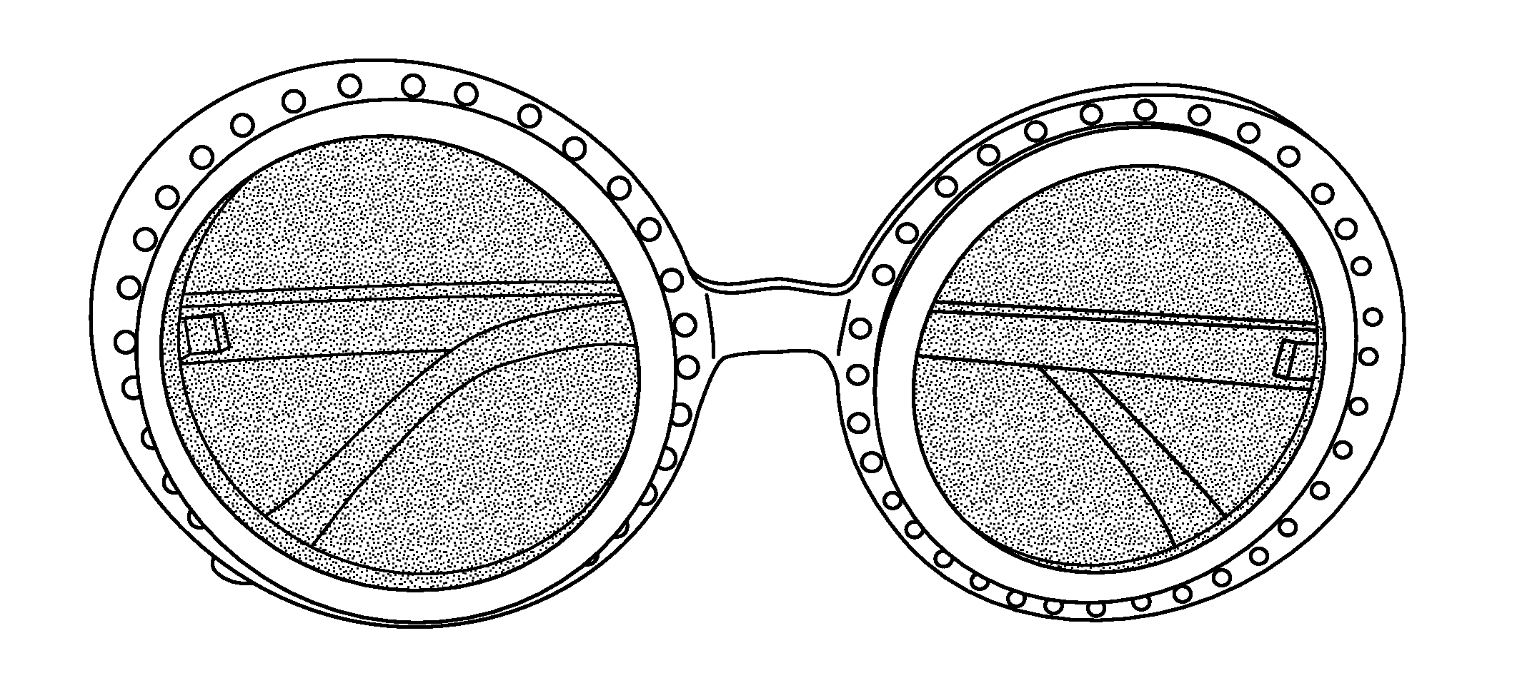
[0108]Illumination devices are based upon high intensity blue light-emitting diodes (LEDs) and emit an intense band of light that is centered at 480 nm and has a half-height bandwidth of about 90 nm. The half-height bandwidth is narrowed to about 70 nm with truncation of a higher-wavelength tail by placing a Wratten #47 filter in front of the focusing and collimating lens of the illuminator housing. The blue light emitted by this configuration is ideally suited for the transdermal excitation of fluorescein and can be effectively blocked by specially-selected yellow-orange lenses that can be mounted in photographic filter holders for photographic documentation purposes or in eyeglass frames for wear by the surgeon or the user.

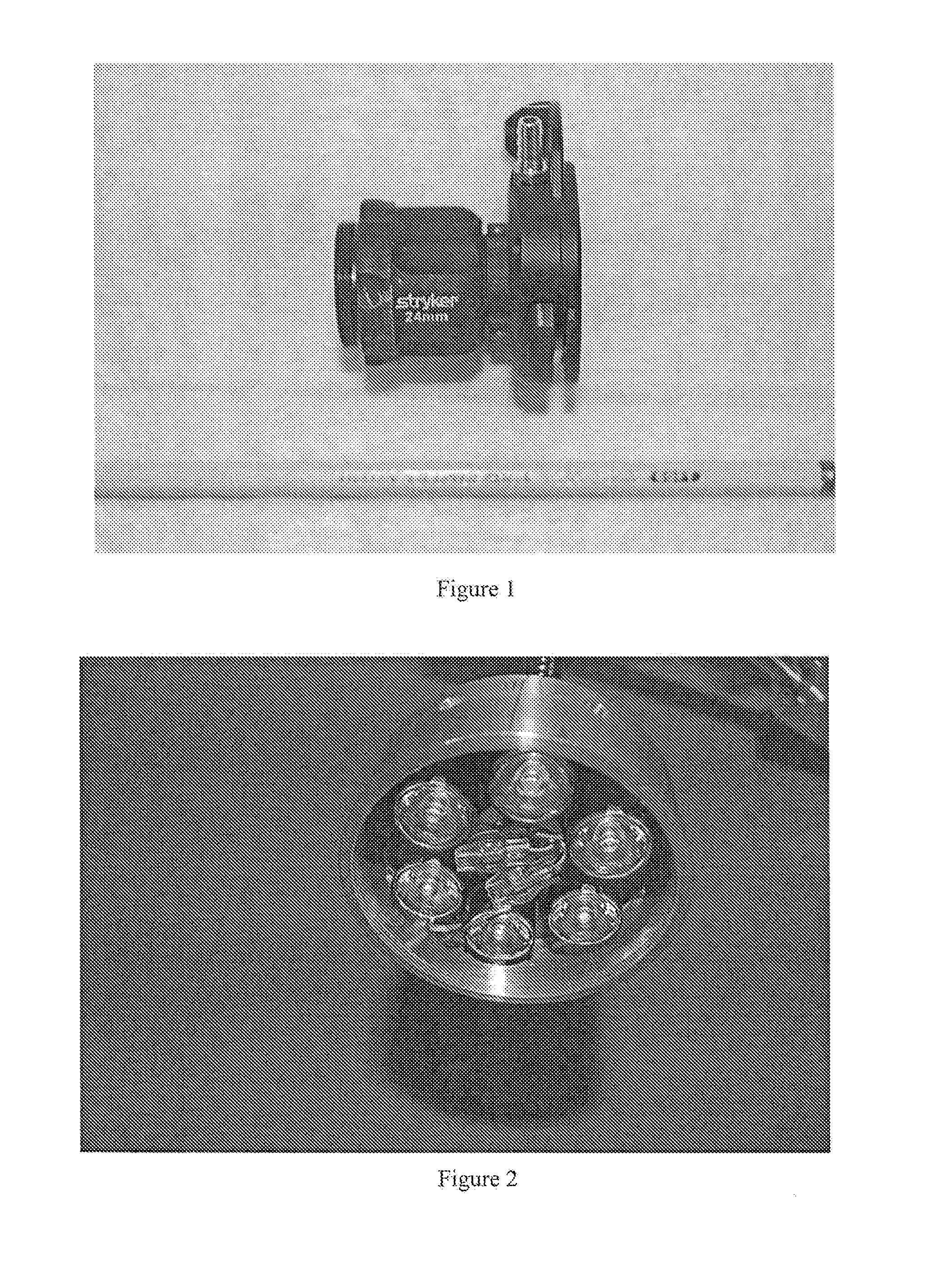
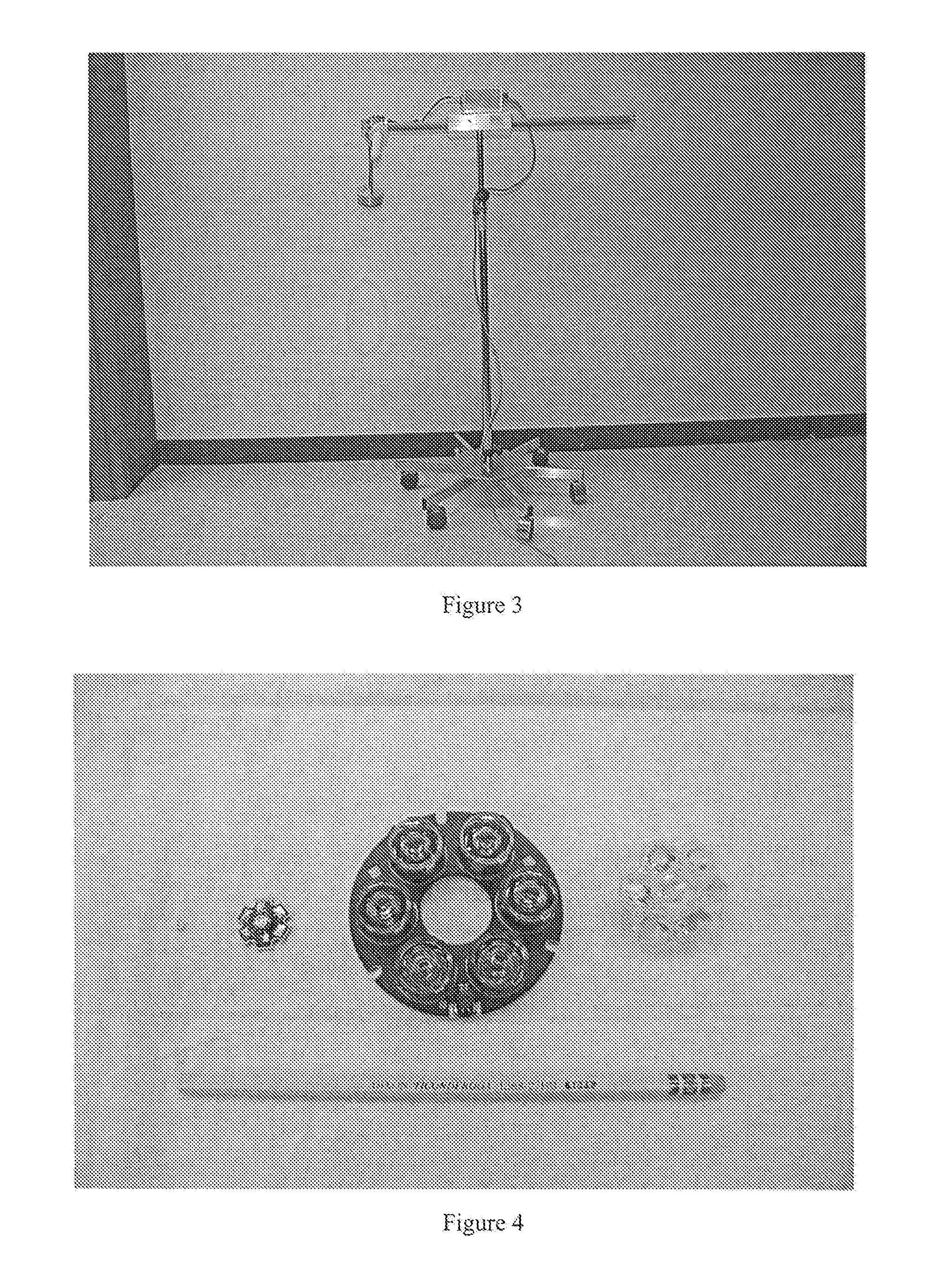
[0109]A holographic notch filter (Kaiser Optical) to remove the intense, but spectrally narrow band of blue light from the LED light sources, was fitted into a surgical telescope camera adapter. The camera adapter design used by Stryker Endoscopy ...

example 2

Materials and Methods

[0113]Fluorescein: 10% Fluorescein USP (Mallinckrodt Baker, Inc., Phillipsburg, N.J.) was diluted in normal saline to concentrations of 0.001%, 0.01%, and 0.1%. The diluted fluorescein was injected (0.5-1.0 ml) into the dermis of the distal forelimb and hindlimb of swine using a 1.0 ml tuberculin syringe. The dose and number of dermal injections varied according to the result. Sometimes the first injection illuminated a large lymphatic trunk easily visible through the skin. This happened more often in the hindlimb than the forelimb. Occasionally, several injections were needed to illuminate a channel. And sometimes, no lymphatic channel filled from the dermal injection. This happened in forelimbs only. Most often, after less than three injections, a lymphatic channel drained from the limb to nodes as seen through the skin and led to the nodal basin containing the sentinel node. In this model, the sentinel nodes were not visible through the skin. An open surgical...

example 3

Lymphatic Mapping

[0116]A total of five swine were studied utilizing all four limbs; the total number of lymphatic nodal basins evaluated was 20. Of the ten fluorescein hindlimb injections, nine (90%) showed lymphatic trunks under the skin leading to the SLN. In the forelimbs available for study, seven fluorescein injections were performed and only one (14%) developed visible fluorescence leading to a SLN. Because of the poor visualization of lymphatic trunks with fluorescein in the forelimbs of swine, we stopped injecting forelimbs in the last animals studied. In the forelimbs, lymphazurin was injected four times and failed to lead to a SLN. We feel that the forelimb lymphatic drainage in the swine may lead to the deep lymphatics more often than the superficial ones as is seen reliably in the swine hindlimb (Wallace, et al. “Lymphoseek: a molecular imaging agent for melanoma sentinel lymph node mapping.” Annals of Surgical Oncology (2007) 14(2):913-21.).

[0117]Injection method: We fo...


PUM


Abstract

The present invention relates generally to the field of fluorescent dyes as a system useful for surgery imaging. More particularly, the present invention relates to systems, methods and kits for exciting fluorescent, phosphorescent or luminescent molecules with light from a light source and detecting the relative fluorescent, phosphorescent, or luminescent light intensity emitted from the fluorescent, phosphorescent, or luminescent molecule. Such systems may be applied as mapping agents for various surgical techniques, such as for cancer surgeries and biopsies.

Description

CROSS-REFERENCE TO RELATED APPLICATION[0001]The present application claims priority to U.S. provisional patent application Ser. No. 61 / 171,414 filed on 21 Apr. 2009, incorporated herein by reference.BACKGROUND OF THE INVENTION[0002]The present invention relates generally to the field of fluorescent dyes as a system useful for surgery imaging. More particularly, the present invention relates to systems, methods and kits for exciting fluorescent, phosphorescent or luminescent molecules with light from a light source and detecting the relative fluorescent, phosphorescent, or luminescent light intensity emitted from the fluorescent, phosphorescent, or luminescent molecule. Such systems may be applied as mapping agents for various surgical techniques, such as for cancer surgeries and biopsies.[0003]The publications and other materials used herein to illuminate the background of the invention or provide additional details respecting the practice, are incorporated by reference.[0004]Signif...

Claims


Application Information

Patent Timeline
Patent Type & Authority Applications(United States)
IPC IPC(8): A61B6/00
CPCA61B5/0059A61B5/0091A61B5/415A61B5/418G01N2201/062A61B2019/5441G01N21/6428G01N21/6447G01N2021/6471A61B19/54A61B90/39A61B2090/3941
Inventor GRISSOM, CHARLES B.MCGREEVY, JAMES M.ANDTBACKA, ROBERT HANS INGEMAR
Owner UNIV OF UTAH RES FOUND
Why Eureka
Try Eureka