
Anti-human crp antibody and its application
A technology for antibody and human detection, applied in the direction of application, anti-animal/human immunoglobulin, peptide, etc., can solve the problem of insufficient risk prediction, and achieve the effect of easy operation and convenient mass production
Patent Information
- Authority / Receiving Office
- CN · China
- Patent Type
- Patents(China)
- Current Assignee / Owner
- Publication Date
- 2018-11-30
Smart Images
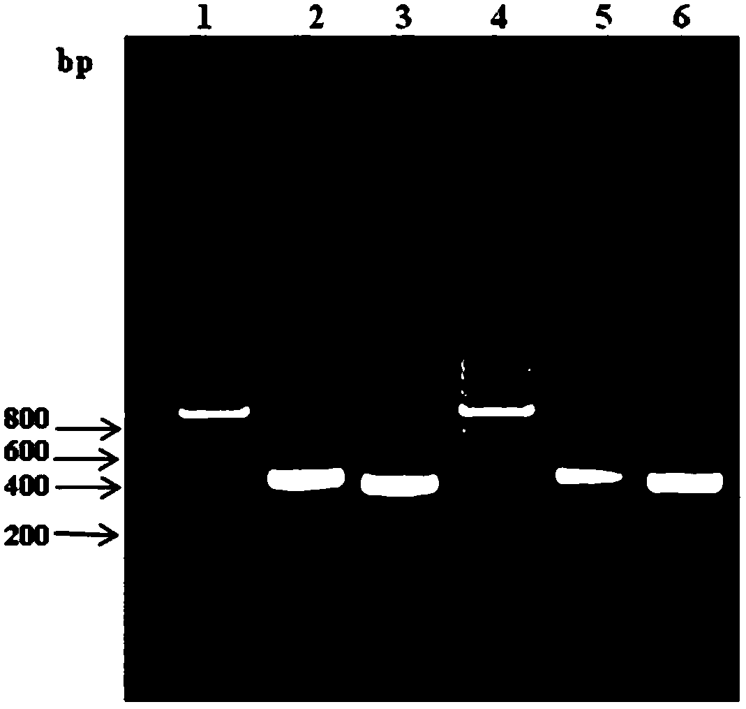
Figure 1 
Figure 2 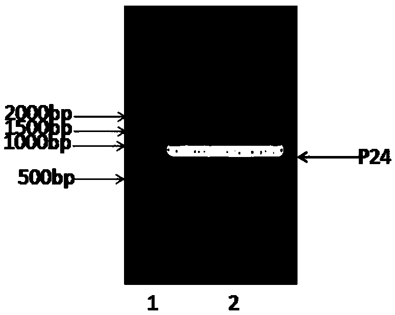
Figure 3
Abstract
Description
technical field
[0001] The invention belongs to the field of biotechnology, and in particular relates to two anti-human C-reactive protein (CRP) antibodies and preparation methods thereof, as well as the application of the antibodies in the detection of human C-reactive protein. Background technique
[0002] C-reactive protein (CRP), a member of the protein family, is an acute phase reactive protein whose plasma concentration rises sharply during tissue damage and bacterial infection. It is an important defense molecule in the body and is mainly produced by the liver. produced and secreted. CRP is composed of five identical spherical monomers non-covalently bonded to form a stable disc-like structure and belongs to the pentamer family. CRP is a symmetrical pentahedron in structure, its monomer is composed of 206 amino acids, its molecular weight is about 23KDa, and the total molecular weight of CRP is about 118KDa. CRP is involved in various inflammatory and immune respons...
Examples
Embodiment 1
[0042] Example 1. Preparation of anti-human C-reactive protein hybridoma cell lines
[0043] 1. Animal Immunization
[0044] BALB / c female mice (purchased from Changzhou Cavens Laboratory Animal Co., Ltd.) were immunized with C-reactive protein extracted from human plasma (purchased from HyTest Company) according to the general immunization procedure. For specific immunization conditions, please refer to the "Experimental Guide for Antibody Preparation and Use". The serum titer of the immunized mice was tracked by indirect ELISA, and the immunized mouse with the highest serum titer was selected for fusion experiments of mouse spleen cells and mouse myeloma cells.
[0045] 2. Cell fusion
[0046] (1). Preparation of spleen cells
[0047] The immunized mice were removed from the eyeballs and blood was collected. After they were sacrificed by cervical vertebra, they were soaked in 75% (v / v) alcohol for 10 minutes. The spleen was removed from the sterile operating table, placed...
Embodiment 2
[0057] Example 2. Determination of antibody variable region sequences of hybridoma cell lines
[0058] The variable region sequences of the hybridoma cell lines M24 and M03 were determined.
[0059] a. RNA extraction: The total RNA of the above hybridoma cell lines M24 and M03 was extracted with reference to the instructions of the cell total RNA extraction kit (purchased from Roche Company) and reverse transcribed immediately;
[0060] b. RNA is reverse transcribed into DNA: with reference to Thermo Scientific Reverted First strand cDNA Synthesis Kit (purchased from Thermo), the total RNA extracted in the previous step was reverse transcribed to obtain cDNA, which was frozen at -20°C for later use;
[0061] c. PCR amplification and recovery of variable region sequences: The cDNA obtained in the previous step was used as a template, and the variable region sequences of the heavy chain and light chain were analyzed using the universal primers for variable region sequences of mo...
Embodiment 3
[0065] Example 3. Recombinant expression and purification of single-chain antibody
[0066] According to the sequencing results in Example 2, a linking peptide (GGGGS) was added between the variable regions of the heavy and light chains of the hybridoma cell lines M24 and M03 antibodies, respectively. 3 , introduced six histidine tags SEQ ID NO: 17, and fused its whole gene with histidine tags to perform codon optimization according to the preference of the Pichia pastoris expression system to carry out the recombinant expression of the single-chain antibody. The expressed antibodies are named as antibody P24 and antibody P03, respectively, and their structural compositions are shown in the appendix. figure 2 shown. The recombinant expression of the above-mentioned single-chain antibody is as follows:
[0067] a) Construction of expression plasmid of fusion protein gene
[0068] The nucleotide sequence of the codon-optimized antibody P24 is shown in SEQ ID NO: 18, and the ...