
Magnetic resonance imaging t2 contrast medium for cell contrasting, and method for manufacturing same
a magnetic resonance imaging and contrast medium technology, applied in the field of magnetic resonance imaging t2 contrast agent, can solve the problems of limited ultrasensitive in vivo tracking of a small number of labeled cells, low relaxivity of spios, poor crystallinity, etc., and achieve the effect of effectively taking up into a cell and effectively labeling various types of cells
Patent Information
- Authority / Receiving Office
- US · United States
- Patent Type
- Applications(United States)
- Current Assignee / Owner
- Publication Date
- 2014-06-19
- Estimated Expiration
- Not applicable · inactive patent
Smart Images
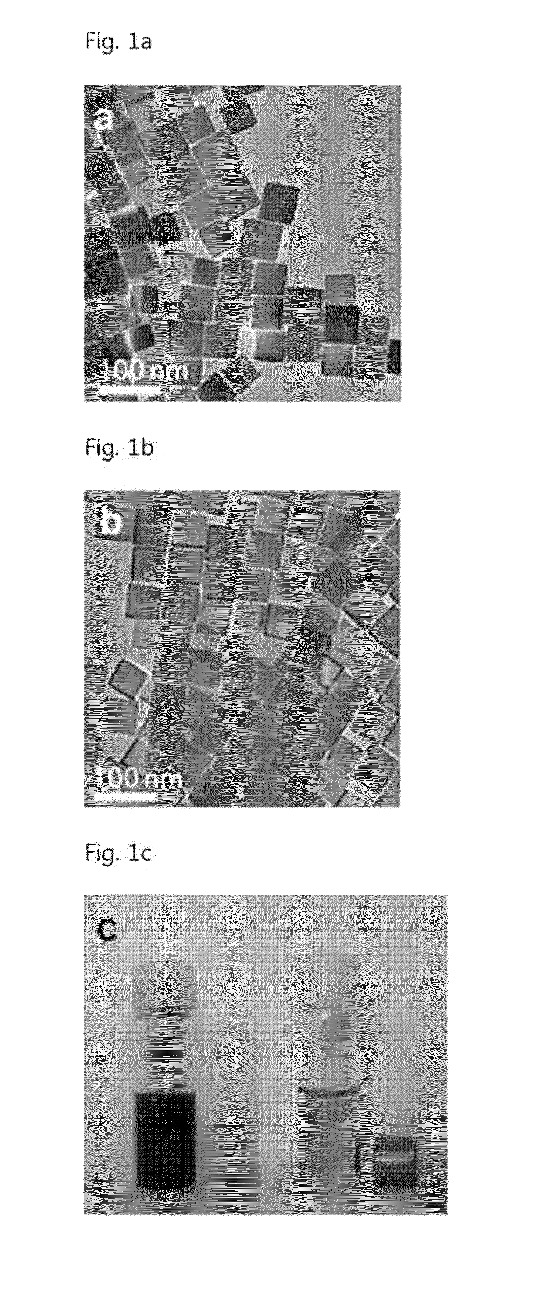
Figure 1 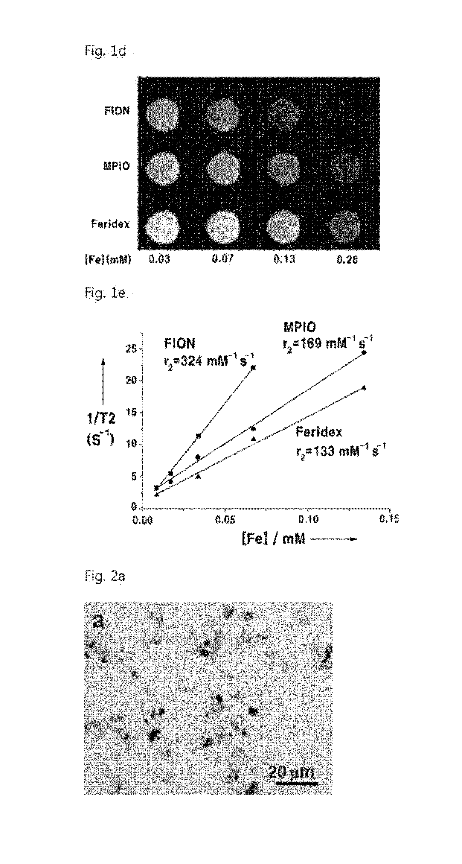
Figure 2 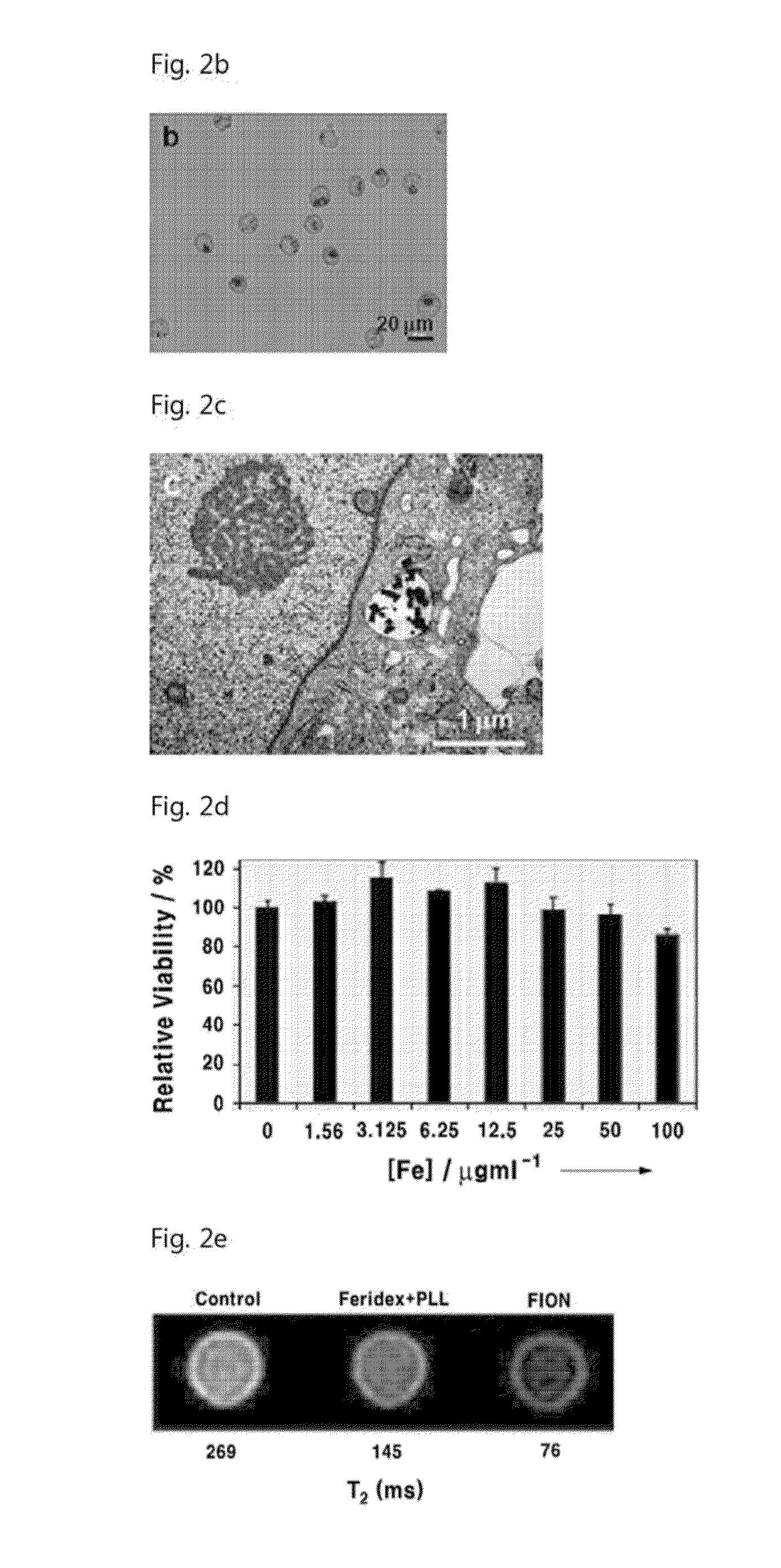
Figure 3
Abstract
Description
TECHNICAL FIELD
[0001] The present invention relates to a magnetic resonance imaging T2 contrast agent (medium) and a method for manufacturing the same, more particularly, a magnetic resonance imaging T2 contrast agent comprising magnetic nanoparticles.BACKGROUND AR
[0002] Magnetic nanoparticles have attracted much attention for a variety of biomedical applications including MRI, drug delivery, hyperthermia, bioseparation, etc.
[0003] Particularly, superparamagnetic iron oxide nanoparticles (SPIOs) such as Feridex, Resovist, etc. have been recently used as a MRI T2 contrast agent since the T2 relaxation time of water adjacent to the nanoparticles is significantly decreased due to high magnetic moment of the nanoparticles. It is possible to increase tiny difference of contrast between tissues by using SPIOs. However, SPIOs generally have relatively low relaxivities since they are synthesized in aqueous media and consequently have poor crystallinity. For ultrasensitive magnetic resonance ...
Examples
example 1
Synthesis of Ferrimmagnetic Iron Oxide Nanocube (FION)
[0073]Iron(II) acetylacetonate was added to a mixture composed of oleic acid and benzyl ether. Remaining air was removed by reducing the mixture solution via a vacuum pump. Then, the mixture solution was heated up to the boiling temperature of benzyl ether, while stirring the mixture solution. The mixture solution was maintained at the boiling temperature for 30 minutes and was cooled in air. Then, a mixture of toluene and hexane was added to the mixture solution, thereafter particles were separated by centrifugation. The separated particles were stored in chloroform.
[0074]In order to render the magnetic nanoparticles hydrophilic and biocompatible, the synthesized magnetic nanoparticles were mixed with 1,2-distearoyl-sn-glycero-3phosphoethanolamine-N-[methoxy(polyethylene glycol)-20001](mPEG-2000 PE, Avanti Polar Lipids, Inc.) in chloroform. After removing chloroform at 80° C., the nanoparticles were dispersed by adding water. E...
example 2
Measurement the Magnetic Resonance Imaging Ability of FION
[0076]In order to measure the magnetic resonance imaging ability, various concentrations of the nanoparticles which were synthesized in Example 1, were dispersed in 1% agarose solution. In order to measure T2 relaxation time of the nanoparticles, T2-weighted images were obtained by using a 1.5T magnetic resonance scanner Also, T2 relaxation time was measured from change of signal intensity with change of TE value, by using the Levenberg-Marquardt algorithm.
[0077]Since the synthesized nanoparticies have strong contrast effect, they show more distinguished contrast effect in T2-weighted images than the conventional superparamagnetic particles (FIG. 1d). As shown in FIG. 1e, the relaxivity of the FIONs was 2 or 3 times higher than that of Feridex which is currently commercialized.
example 3
Cellular Uptake of the Magnetic Nanoparticles
[0078]For cellular uptake of the magnetic nanoparticles synthesized in Example 1, the nanoparticles were cultured for 24 hours together with the prepared cells. The cells were detached from the culture plate by trypsin treatment, in order to remove the nanoparticles which were not taken up. Ficoll-paque was placed in the centrifugation tube, after then the cellular dispersion solution was added carefully on the Ficoll-paque layer, thereby separation between the cellular dispersion solution layer and the Ficoll-paque layer. When the centrifugation was performed at this state, the nanoparticles with high density were settled down under the Ficoll-paque layer, while the cells with low density were floated on the Ficoll-paque layer.
[0079]In order to stain the intracellular magnetic nanoparticles, the cells from which unabsorbed nanoparticles were removed were cultured in an 8-well chamber slide and were fixed with 4% paraformaldehyde. The iro...