
Method of detection of DNA end(s) and its use
Patent Information
- Authority / Receiving Office
- US · United States
- Current Assignee / Owner
- Publication Date
- 2020-08-13
Smart Images
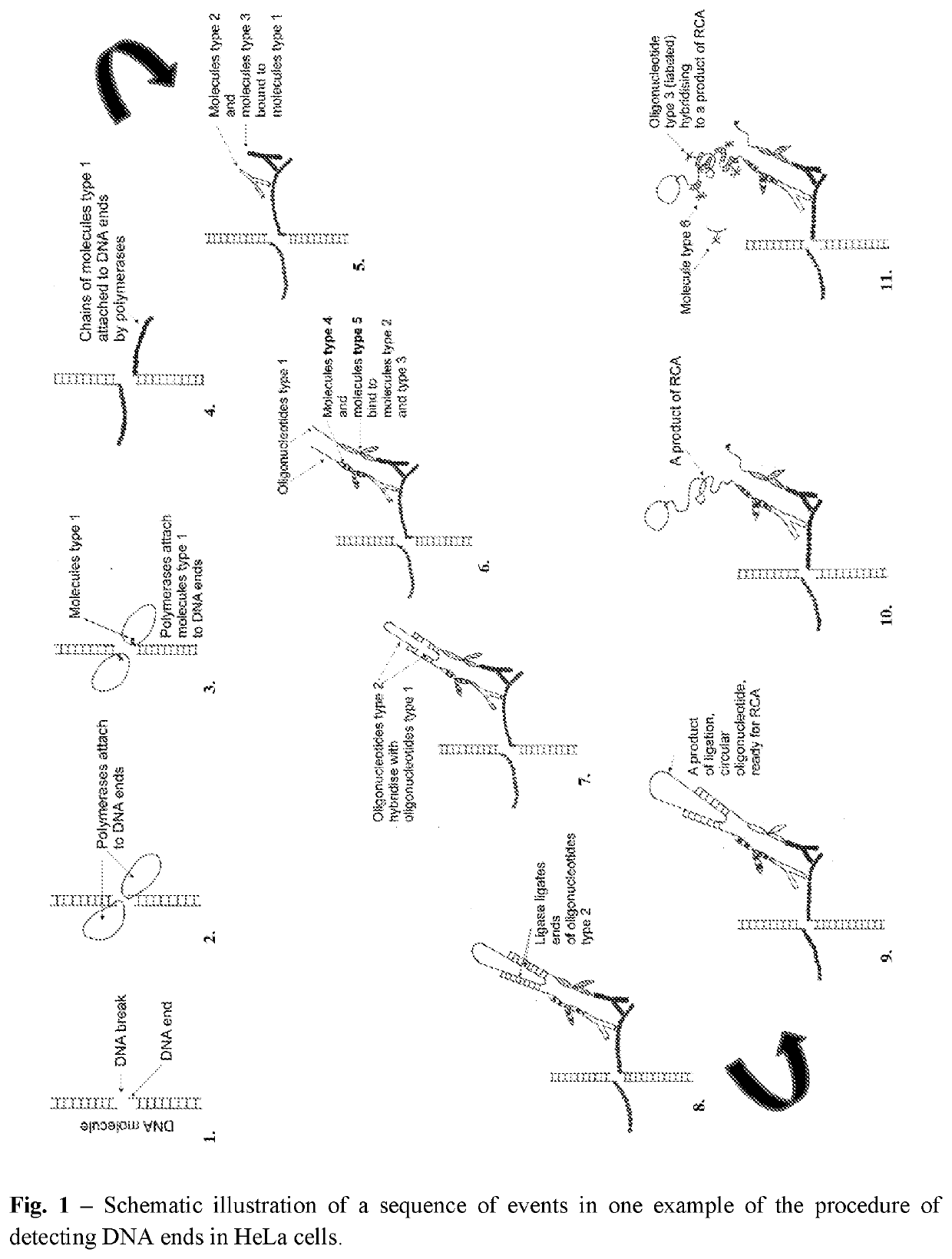
Figure 1 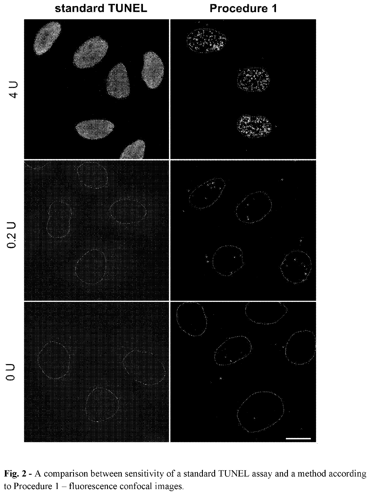
Figure 2 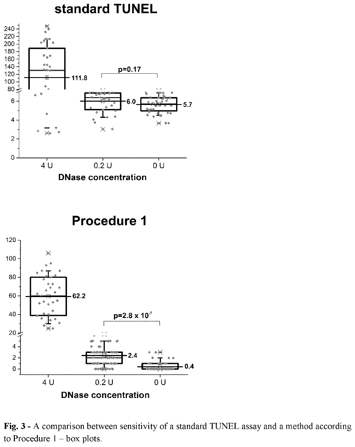
Figure 3
Abstract
Description
FIELD OF THE INVENTION
[0001] The presented invention falls within the molecular diagnostics and biomedical field. It is related to the method of detection and / or quantitative and / or qualitative analysis of DNA damage. More precisely, the present invention enables direct, highly sensitive, specific and efficient recognition and / or detection of different types of free DNA ends formed in result of induction of single- or double-strand DNA breaks in the cell nucleus, or in isolated biological materials.BACKGROUND
[0002] For a life to continue from generation to generation, genomes of organisms must be passed on without any significant changes. The storage of genetic information in a form of a unique sequence of nucleobases that form deoxyribonucleic acid (DNA), and the ability to transfer it through generations, are principal features of all living organisms. Cells are the basic structural and functional units of living organisms. The sequence of bases must, therefore, be preserved during ...
Examples
example 1
[0370]Detection of Free DNA Ends in Untreated and DNase I Treated Fixed HeLa Cells Using the Method According to Procedure 1.
[0371]In this experiment HeLa 21-4 cells (obtained from P.R. Cook, University of Oxford) were used. Cells were seeded on 18-mm coverslips (number of cells: 0.2×10⁶ per 1 coverslip) and cultured for 3 days in DMEM supplemented with 10% FBS. Subsequently, cells were incubated in 70% ethanol in water for 12 hours at −20° C. Then, cells were incubated in 0.5 mM EDTA in water for 30 minutes at room temperature. Before processing of DNA ends, some samples were treated with DNase I in order to induce DNA breaks. The reaction was performed in a droplet for 30 minutes at room temperature in a reaction mixture consisting of: 0.2 or 4 units of DNase I (Thermo Fisher Scientific, AM2222), 1× DNase I buffer (Thermo Fisher Scientific, AM8170G) and water. Subsequently, BrdU was linked to free ends of DNA using TdT enzyme using APO-BRDU kit (Phoenix Flow Systems, AU: 1001). Th...
example 2
[0381]Detection of Free DNA Ends in Untreated Fixed HeLa Cells Using the Method According to Procedure 2
[0382]In this experiment HeLa 21-4 cells (obtained from P.R. Cook, University of Oxford) were used. The cells were seeded on 18-mm coverslips (number of cells: 0.2×10⁶ per 1 coverslip), cultured for 3 days in DMEM supplemented with 10% FBS and then incubated in 70% ethanol in water for 12 hours at −20° C. After that the samples were treated according to step 3 of Procedure 2 (blocking endogenous biotin) (Molecular Probes, Endogenous Biotin Blocking Kit, E-21390). Increasing of accessibility of DNA ends (step 4) was performed with 0.5 mM EDTA in water for 30 min at RT. To incorporate unmodified and modified biotin-conjugated nucleotides, cells were dipped in UltraPure Distilled Water (Invitrogen, Thermo Fisher Scientific, 10977-035) and then incubated (1 hour incubation at 37° C. in a humid chamber) with reaction mixture consisting of 1× NEBuffer2 (NEBiolabs, B7002S), 30 μM of each...
example 3
[0385]Detection of Free DNA Ends in Fixed HeLa Cells Using the Method According to Procedure 2—Evaluation of the Effect of Blocking Endogenous Biotin.
[0386]HeLa 21-4 cells were treated as in Example 2 but without step 3 (pre-blocking) from Procedure 2.
[0387]Results:
[0388]Free DNA ends, represented as bright fluorescent foci, were readily detected in untreated HeLa cells processed according to Procedure 2 (without step 3—blocking endogenous biotin) (FIG. 4., second row, left image). The average number of foci detected per one nucleus was 128±6 (N=24).
[0389]Conclusion (Example 2 and 3):
[0390]Slightly more free DNA ends were detected in samples processed according to Procedure 2 without blocking endogenous biotin (step 3) than in samples in which this step was present (128±6 vs 116±6). However, an independent two-sample t-test yielded a p-value=0.15, indicating that the difference between these two samples is not statistically significant. Thus, one can conclude that blocking endogenou...