Image processing process based on magnetic resonance three-dimensional renogram

An image processing and magnetic resonance technology, applied in image data processing, magnetic resonance measurement, image analysis, etc., can solve problems such as difficulty in providing valuable clinical information, unacceptable workload time, and large nephrogram errors, achieving MRR Accurate, time-saving, and work-intensive effects

Inactive Publication Date: 2008-03-12
PEKING UNIV
View PDF0 Cites 25 Cited by
  • Summary
  • Abstract
  • Description
  • Claims
  • Application Information

AI Technical Summary

Problems solved by technology

Due to the inability to correct the image movement caused by breathing and to automatically and efficiently segment the cortex and medulla, the error of the renogram is relatively large, and it is difficult to provide valuable clinical information, which brings difficulties to subsequent research.
[0004] At present, doctors can only obtain the region of interest and perform data

Method used


Image

  • Image processing process based on magnetic resonance three-dimensional renogram
  • Image processing process based on magnetic resonance three-dimensional renogram
  • Image processing process based on magnetic resonance three-dimensional renogram

Examples


Embodiment Construction

[0024] The present invention will be described in detail below in conjunction with the accompanying drawings and embodiments.

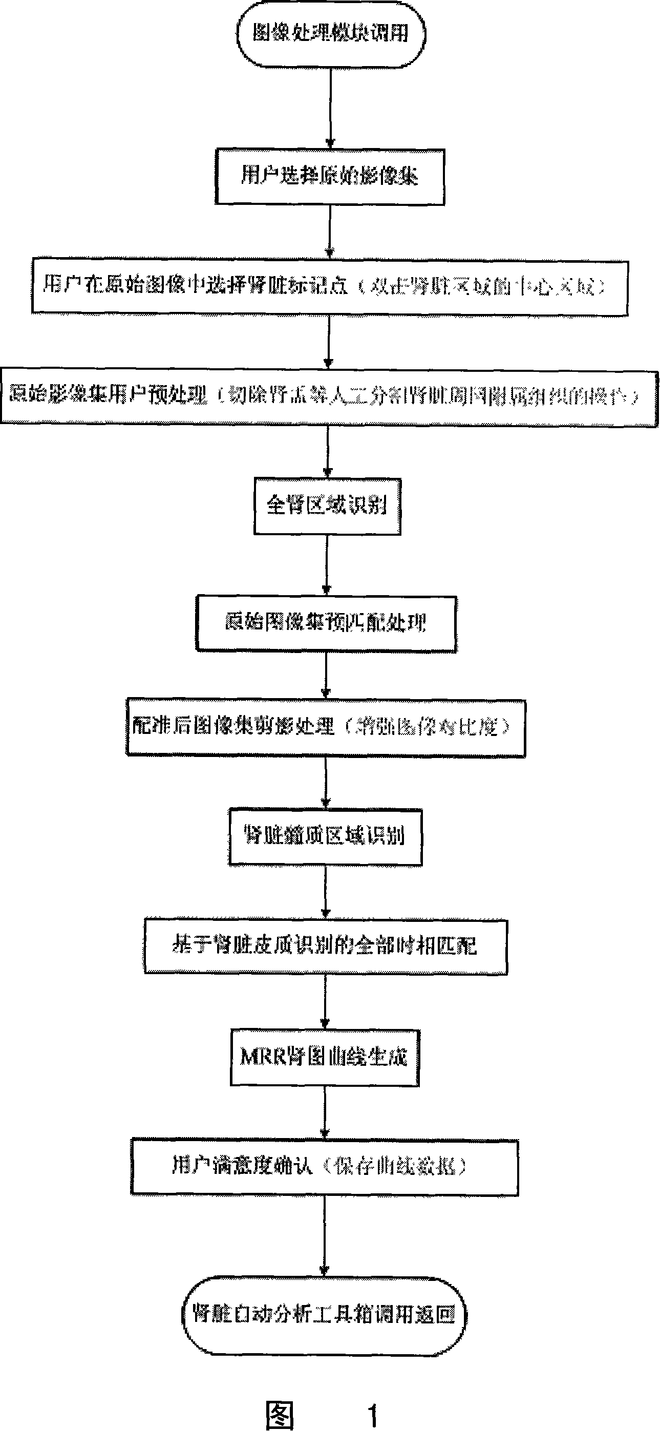
[0025] As shown in Figure 1, an image processing method of a magnetic resonance three-dimensional renogram based on region growth and correlation matching in the present invention, its specific operation process is as follows:

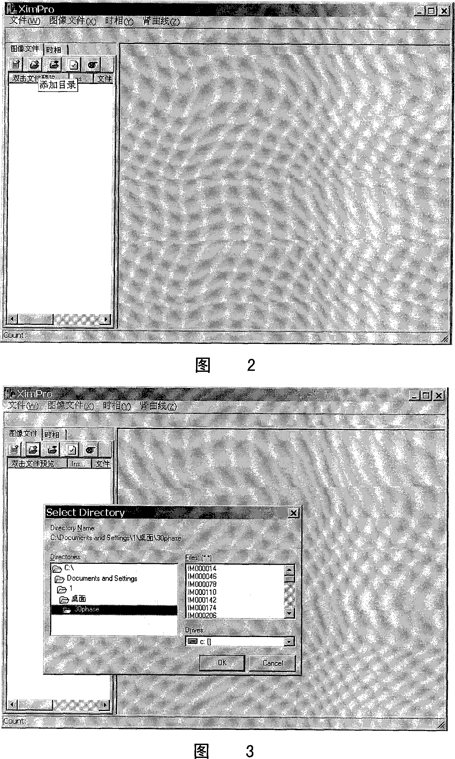
[0026] The first step: run the program, the program interface appears (as shown in Figure 2), call the image processing module, select the image directory to be processed (as shown in Figure 3), read the image, the MRI image obtained from the magnetic resonance workstation It is in DICOM (Digital Imaging Communications in Medicine) format.

[0027] DICOM files refer to medical files stored in accordance with the DICOM standard. The DICOM file header contains relevant information identifying the data set, in other words, a lot of information about the image is stored in the file header. Since the order of filenames of DICOM i...


PUM


Abstract

The present invention relates to an image processing method which bases on a magnetic resonance three-dimensional renogram. The present invention includes the steps mentioned below. Firstly, a magnetic resonance three-dimensional scanning image of kidneys is read from a magnetic resonance workstation. Secondly, every phase image number and background phase, a cortex and medulla demarcation phase and a holonephros phase are ensured. Thirdly, relative vascular and renal pelvis regions in the image of the holonephros phase are excised. Fourthly, a holonephros contour is identified. The fifth step is to match the background phase with the cortex and medulla demarcation phase and silhouette. The sixth step is to segment the medulla and the cortex of the kidneys. The seventh step is to match the images of all phases. The present invention provides a novel kidney dynamic reinforced image segment and match scheme and can precisely correct the motions caused by the breath of the kidneys during the MRI image obtaining process and precisely segment the cortex and the medulla. The image processing method of the present invention is proved by the practices to be capable of obtaining the reasonable three-dimensional magnetic resonance renogram. The present invention can be widely applied to the image processing of the magnetic resonance renogram.

Description

technical field [0001] The invention relates to a magnetic resonance image processing method, in particular to an image processing method based on a magnetic resonance three-dimensional renogram. Background technique [0002] MRI (Magnetic Resonance Imaging, Magnetic Resonance Imaging) dynamic contrast-enhanced scan, as a safe way to evaluate renal function, can be used to evaluate renal perfusion and filtration functions, and has important clinical application prospects. During dynamic contrast-enhanced renal MR (magnetic resonance) scanning, MRI contrast agents pass through the cortex, medulla, and collection system of the kidney in sequence, and the changes in signal intensity of contrast agents in different regions relative to time can reflect some functions of the region. The time-signal intensity curve formed by the change of signal intensity is called MRR (Magnetic Resonance Renography), also called magnetic resonance renography. Magnetic resonance renography, with h...

Claims


Application Information

Patent Timeline
IPC IPC(8): A61B5/055G01R33/44G06T7/00
Inventor 庄杰吕东姣王晶陈慧军许玉峰王霄英张珏方竞
Owner PEKING UNIV
Why Eureka
Try Eureka