Eye ground retina OCT image correction method

An image correction, retinal technology, applied in the field of optoelectronics, can solve the problem that the retina of the fundus cannot be measured by the OCT image of the retina of the rectangular fundus.

Active Publication Date: 2014-11-19
SHENZHEN CERTAINN TECH CO LTD
View PDF8 Cites 18 Cited by
  • Summary
  • Abstract
  • Description
  • Claims
  • Application Information

AI Technical Summary

Problems solved by technology

[0005] The invention provides a correction method after retinal OCT imaging, and its purpose is to solve the defect that the fundus

Method used


Image

  • Eye ground retina OCT image correction method
  • Eye ground retina OCT image correction method
  • Eye ground retina OCT image correction method

Examples


Embodiment Construction

[0064] In order to make the technical problems, technical solutions and beneficial effects to be solved by the present invention clearer, the present invention will be further described in detail below in conjunction with the accompanying drawings and embodiments. It should be understood that the specific embodiments described here are only used to explain the present invention, not to limit the present invention.

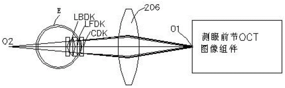
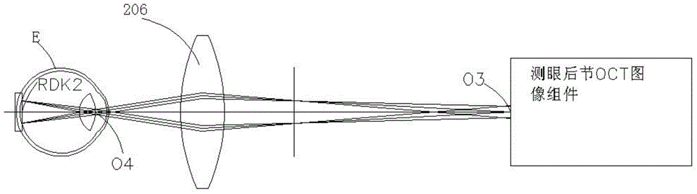
[0065] The present invention mainly describes the restoration of the uncorrected rectangular fundus retinal OCT image processed by a computer into an OCT image of the true shape of the fundus retina. In the restoration process, it is necessary to obtain the center position of the scanning fan-shaped area where the scanning beam scans the fundus retina and the scanning angle uO45. The scanning angle uO45 is obtained according to uO45=γ·uO4. Among them, γ represents the overall angular magnification of the anterior segment, which is calculated according to the cente...


PUM


Abstract

The invention discloses an eye ground retina OCT image correction method. The method comprises the steps that scanning beams scan an anterior segment and a posterior segment, and meanwhile a human anterior segment OCT image and an eye ground retina OCT image are obtained, wherein the human anterior segment OCT image contains a corneal OCT image, a crystalline lens anterior surface OCT image and a crystalline lens posterior surface OCT image, and the human anterior segment OCT image and the eye ground retina OCT image are processed by a computer without correction; the OCT images are corrected and restored to images with a true form to obtain the corneal anterior surface curvature radius, the corneal posterior surface curvature radius, the crystalline lens anterior surface curvature radius, the crystalline lens posterior surface curvature radius, the corneal thickness, the anterior chamber depth and the crystalline lens thickness; a rendezvous point (O45) of center lines of the scanning beams which are refracted by the posterior surface of a crystalline lens is determined, and the circle center of the sector scanning region and the scanning angle (uO45) are calculated; the uncorrected eye ground retina OCT image is restored to an image with a true form, and finally a true sectional image of an eye ground retina is obtained through restoration and the curvature of the eye ground retina is measured according to a measured anterior segment OCT image and a measured posterior segment OCT image.

Description

technical field [0001] The invention belongs to the field of optoelectronic technology, and in particular relates to a method for correcting fundus retinal OCT images. Background technique [0002] Nowadays, many ophthalmic diagnostic instruments are used to measure many parameters of the human eye to help doctors diagnose patients' eye diseases. However, in the prior art, there is no accurate, non-destructive and reliable measurement method for the curvature of the front and rear surfaces of the human eye lens and the curvature of the retina of the human eye fundus. [0003] The current OCT technology is divided into three types: time domain, frequency domain and frequency sweep. Among them, the time-domain OCT technology has a deep detection depth, but the speed is too slow. After the measured human eye moves, it will affect the tomographic image of the human eye. Tomography of the eye. If many structural parameters of the human eye cannot be measured at the same time, ...

Claims


Application Information

Patent Timeline
IPC IPC(8): A61B3/12A61B3/14
Inventor 蔡守东吴蕾
Owner SHENZHEN CERTAINN TECH CO LTD
Why Eureka
Try Eureka