

Method applied to quantitative detection of carbon dioxide exhaled by cancer cells
A technology for quantitative detection of carbon dioxide, applied in the field of fluorescent biosensors, can solve problems such as the lack of quantitative detection of cellular carbon dioxide and the inability to monitor the metabolic process of cancer cells
- Summary
- Abstract
- Description
- Claims
- Application Information
AI Technical Summary
Problems solved by technology
Method used
Image

Examples
Embodiment 1
[0043] (1) Dissolve 0.64mg TPP-DMAE in 0.01mL dimethyl sulfoxide (DMSO) to obtain a concentration of 1×10 -2 mol / L solution a; add 10mL high-sugar DMEM medium to solution a to obtain a concentration of 1×10 -5 mol / L solution b.
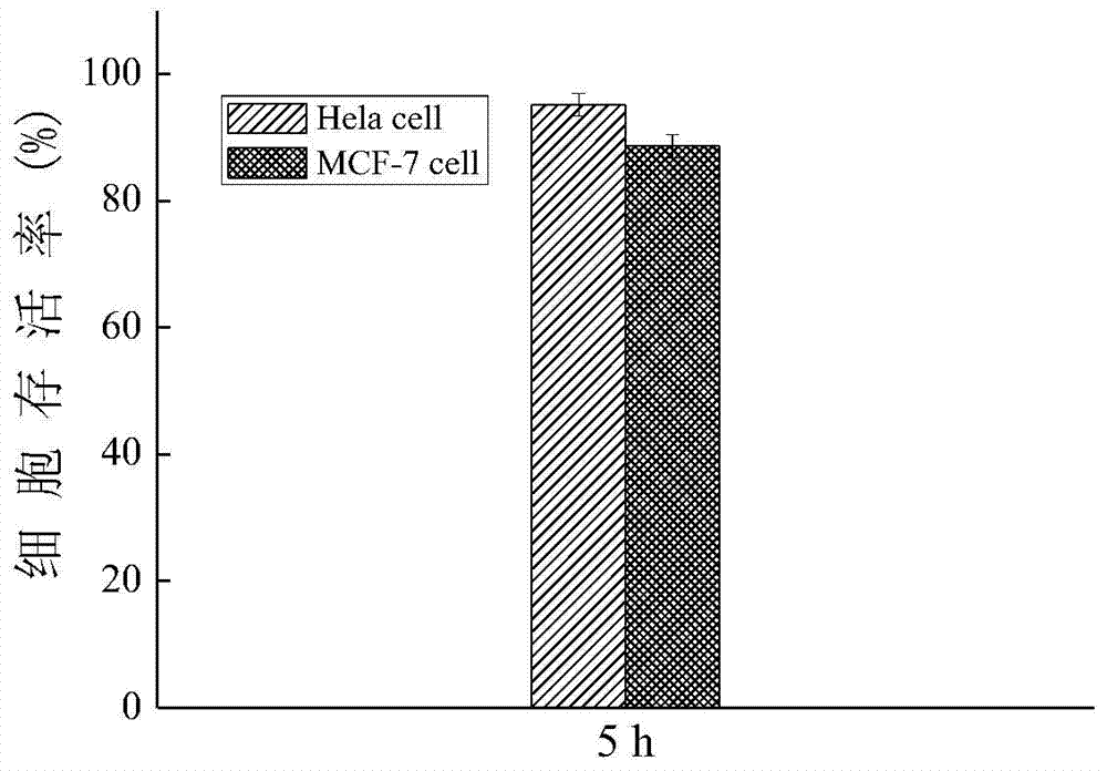
[0044] (2) ① In order to detect whether TPP-DMAE can cause the inactivation of Hela cell and MCF-7cell within the test time range, the following toxicity test experiments were done:
[0045] Cervical cancer cells (Hela cells) and breast cancer cells (MCF-7 cells) were respectively passaged into two culture dishes dedicated to confocal laser microscopy (confocal); the Hela cells, MCF-7cell was digested with 0.05% trypsin until 50-60% of the cells were detached from the bottom of the culture dish, and 3 mL of high-sugar DMEM medium was added to the culture dish of Hela cell and MCF-7 cell to stop the digestion, and then Hela cell Cell and MCF-7cell suspensions were transferred from the petri dish to two 15mL centrifuge tubes, each supplemented with 7m...
Embodiment 2
[0055] (1) Dissolve 0.64mgTPP-DMAE in 0.01mL dimethyl sulfoxide (DMSO) to obtain a concentration of 1×10 -2 mol / L solution a; add 10mL high-sugar DMEM medium to solution a to obtain a concentration of 1×10 -5 mol / L solution b.
[0056] (2) ① In order to detect whether TPP-DMAE can cause the inactivation of Hela cell and MCF-7cell within the test time range, the following toxicity test experiments were done:
[0057] Cervical cancer cells (Hela cells) and breast cancer cells (MCF-7 cells) were respectively passaged into two culture dishes dedicated to confocal laser microscopy (confocal); the Hela cells, MCF-7cell was digested with 0.05% trypsin until 50-60% of the cells were detached from the bottom of the culture dish, and 3 mL of high-sugar DMEM medium was added to the culture dish of Hela cell and MCF-7 cell to stop the digestion, and then Hela cell Cell and MCF-7cell suspensions were transferred from the petri dish to two 15mL centrifuge tubes, each supplemented with 7mL...
Embodiment 3
[0067] (1) Dissolve 0.64mg TPP-DMAE in 0.01mL dimethyl sulfoxide (DMSO) to obtain a concentration of 1×10 -2 mol / L solution a; add 10mL high-sugar DMEM medium to solution a to obtain a concentration of 1×10 -5 mol / L solution b.
[0068] (2) ① In order to detect whether TPP-DMAE can cause the inactivation of Hela cell and MCF-7cell within the test time range, the following toxicity test experiments were done:
[0069] Cervical cancer cells (Hela cells) and breast cancer cells (MCF-7 cells) were respectively passaged into two culture dishes dedicated to confocal laser microscopy (confocal); the Hela cells, MCF-7cell was digested with 0.05% trypsin until 50-60% of the cells were detached from the bottom of the culture dish, and 3 mL of high-sugar DMEM medium was added to the culture dish of Hela cell and MCF-7 cell to stop the digestion, and then Hela cell Cell and MCF-7cell suspensions were transferred from the petri dish to two 15mL centrifuge tubes, each supplemented with 7m...
PUM
 Abstract
Description
Claims
Application Information