Lung tissue single-channel fixing instrument

A single-channel, fixed instrument technology, applied in the direction of sampling devices, etc., can solve the problems of long exposure time of lung tissue, tissue miniaturization, and accelerate the air discharge of lung tissue, and achieve the effect of facilitating immunohistochemical and pathological analysis and fixing integrity

Active Publication Date: 2016-03-09
ZHEJIANG CHINESE MEDICAL UNIVERSITY
View PDF7 Cites 3 Cited by
  • Summary
  • Abstract
  • Description
  • Claims
  • Application Information

AI Technical Summary

Problems solved by technology

[0005] The present invention solves the defects that the existing lung tissue fixation speed is slow, affects the immersion efficiency of the fixative solution, causes the exposure time of the lung tissue to be too long, and affects the experimental results, and provides a lung tissue single-channel fixation instrument, in which an air core is set in the single channel , extract the air inside the fixer through air core pulling, accelerate the discharge of air inside the lung tissue, and also speed up the immersion of the fixative solution, shorten the exposure time of the lung tissue, and ensure the correctness of the experimental results
[0006] The present invention also solves the defects of severe tissue shrinkage caused by the existing lung tissue fixation, which easily destroys the normal structure of the lung tissue and affects the experimental results, and provides a single-channel fixation instrument for lung tissue. The core accelerates the discharge of air inside the lung tissue, accelerates the immersion of the fixative, does not damage the normal structure of the lung tissue, and ensures the correctness of the experimental results

Method used


Image

  • Lung tissue single-channel fixing instrument
  • Lung tissue single-channel fixing instrument
  • Lung tissue single-channel fixing instrument

Examples


Embodiment 1

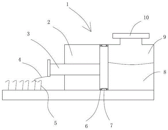
[0021] Embodiment 1: A kind of lung tissue single-channel fixation instrument (see figure 1 ), including a fixed body 1, the fixed body is cylindrical, the fixed body is provided with a containing cavity 9 containing the fixed liquid 8, the upper part of the fixed body is provided with an opening communicating with the containing cavity, and the opening is provided with a sealing cover 10, the fixed body The side wall is provided with a side channel 2 communicating with the containing cavity, and an air core 3 is provided in the side channel.

[0022] The height of the side passage is equal to the height of the accommodation cavity, that is, the air core pull directly divides the inside of the fixed body into the accommodation chamber and the side passage. The air core is in the shape of a piston, including a cylindrical piston head and a piston rod. The piston head is in the side channel, and the piston rod protrudes out of the side channel. The piston head is disc-shaped, a...

Embodiment 2

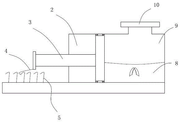
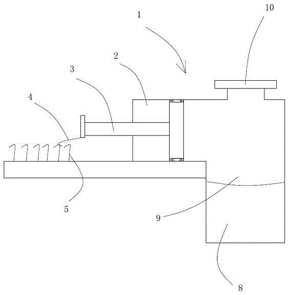
[0024] Embodiment 2: A kind of lung tissue single-channel fixation instrument (see image 3 Figure 4 ), the difference from Embodiment 1 is that the height of the side channel is lower than that of the housing cavity, the side channel is at the upper part of the fixed body, and the liquid level of the fixative is lower than the lower side of the side channel. Refer to Example 1 for all the other structures.

Embodiment 3

[0025] Embodiment 3: A lung tissue single-channel fixation instrument, which is different from the above embodiments in that: a hollow expansion body is connected to the surface of the sealing cover facing the accommodating cavity, and the expansion body is filled with air at standard pressure. The expansion coefficient of the expander is equivalent to the elastic recovery coefficient of the lung tissue, and the expansion of the expander occurs after the lung tissue is emptied of air. Refer to embodiment 1 or embodiment 2 for all the other structures.


PUM


Abstract

The invention relates to a lung tissue single-channel fixing instrument and overcomes the defects that existing lung tissue fixing is low in speed, fixing liquid immersion efficiency is affected, the exposure time of lung tissues is excessively long, and the experiment result is affected. The lung tissue single-channel fixing instrument comprises a fixing body, the fixing body is internally provided with a containing cavity for containing fixing liquid, the fixing body is provided with an opening communicated with the containing cavity, a sealing cover is arranged at the opening, the side wall of the fixing body is provided with a side channel communicated with the containing cavity, and the side channel is internally provided with an air pulling core. As the air pulling core moves outwards, the volume of the containing cavity is increased, so that the pressure intensity in the containing cavity is lowered, a negative pressure state is formed, in the negative pressure environment, air in the lung tissues is discharged rapidly, under the elasticity of the lung tissues, the fixing liquid is immersed into pulmonary alveoli rapidly, and the pulmonary alveoli recovers to the original shape, so that lung tissue cells are integrally fixed, and subsequent immunohistochemistry and pathological analysis are facilitated.

Description

technical field [0001] The invention relates to a tissue fixation device, in particular to a lung tissue single-channel fixation instrument. Background technique [0002] When making pathological sections of animal lung tissue, it is necessary to fix the lung tissue. When observing the lung tissue staining, we not only need to observe clear stained tissue, but also need to keep the shape of the lung tissue intact to avoid tissue damage and alveolar collapse. . [0003] Currently, there are three main lung tissue fixation methods: 1. Using embedding cassettes, the lung tissue is fixed in formaldehyde solution. 2. Put a small ball of cotton on the surface of the formaldehyde solution put into the lung tissue to infiltrate the lung tissue. 3. Use an infusion tube to inject a small amount of formaldehyde solution into the lungs through the trachea. [0004] Although method 1 can quickly infiltrate the lung tissue into the formaldehyde solution, it will cause serious lung coll...

Claims


Application Information

Patent Timeline
Patent Type & Authority Applications(China)
IPC IPC(8): G01N1/06
CPCG01N1/06
Inventor 寿旗扬施佳君李园园傅惠英陈民利
Owner ZHEJIANG CHINESE MEDICAL UNIVERSITY
Why Eureka
Try Eureka