PET (positron emission computed tomography)-fluorescence dual-mode intraoperative navigation imaging system and imaging method implemented by same

A fluorescence imaging and imaging system technology, applied in the field of biomedical imaging, can solve the problems of poor tumor location, limited promotion, limited assistant role of doctors, etc., and achieve the effect of convenient operation and many degrees of freedom.

Active Publication Date: 2017-02-22
北京锐视康科技发展有限公司
View PDF10 Cites 5 Cited by
  • Summary
  • Abstract
  • Description
  • Claims
  • Application Information

AI Technical Summary

Problems solved by technology

Most research on PET surgical navigation focuses on how to quickly fuse preoperative PET images and intraoperative computerized tomography CT images to achieve PET / CT on-the-spot registration, but this technology is limited by the surgical part of the patient during the operation. The change of the position of the positron (β+) nuclides can only be guided at the initial stage of the operation, and cannot be guided in real time during the operation; in addition, scientists in the Netherlands and Switzerland used gamma detectors to count and measure positron (β+) nuclides as a method for real-time tumor resection during the operation. However, due to the poor positioning of the tumor site by this technology, the auxiliary effect on the doctor during the operation is limited, and the subsequent further promotion is also limited.

Method used


Image

  • PET (positron emission computed tomography)-fluorescence dual-mode intraoperative navigation imaging system and imaging method implemented by same
  • PET (positron emission computed tomography)-fluorescence dual-mode intraoperative navigation imaging system and imaging method implemented by same
  • PET (positron emission computed tomography)-fluorescence dual-mode intraoperative navigation imaging system and imaging method implemented by same

Examples


Embodiment Construction

[0038] The present invention will be further described through the embodiments below in conjunction with the accompanying drawings.

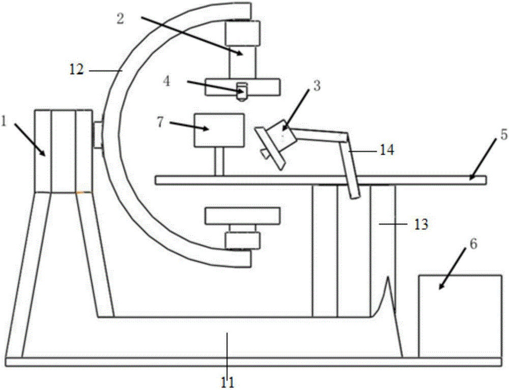
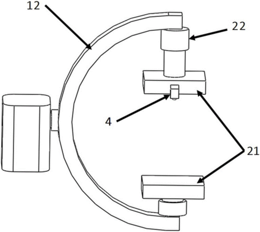
[0039] Such as figure 1 As shown, the PET-fluorescence dual-mode navigation imaging system of this embodiment includes: PET imaging device 2, fluorescence imaging device 3, spatial registration device 4, mechanical control frame 1, imaging bed 5, computer 6 and display device 7; Wherein, the imaging sample is placed on the imaging bed 5, and the imaging bed is installed on the mechanical control frame 1; the PET imaging device 2, the fluorescence imaging device 3 and the spatial registration device 4 are respectively installed on the mechanical control frame 1 and facing the imaging sample; The PET imaging device 2 , the fluorescent imaging device 3 , the spatial registration device 4 , the mechanical control frame 1 and the display device 7 are respectively connected to the computer 6 through data lines.

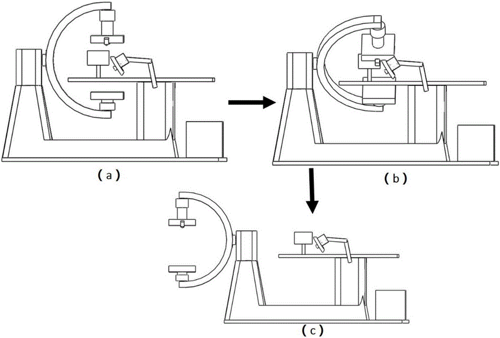
[0040] Such as figure 1 As shown, th...


PUM


Abstract

The invention discloses a PET (positron emission computed tomography)-fluorescence dual-mode intraoperative navigation imaging system and an imaging method implemented by the same. The imaging method includes forming three-dimensional surface contour images of imaging samples by the aid of a spatial registration device; acquiring three-dimensional PET images of internal structures of the imaging samples by the aid of a PET imaging device; carrying out image registration and fusion on the three-dimensional surface contour images and the three-dimensional PET images to obtain three-dimensional fused images of imaging targets; acquiring two-dimensional fluorescence images of the imaging samples in real time by the aid of a fluorescence imaging device; projecting the three-dimensional fused images towards the two-dimensional fluorescence images along the normal axis of an imaging surface of the fluorescence imaging device and displaying depth information of the imaging targets on the two-dimensional fluorescence images. The three-dimensional fused images of the imaging targets comprise surface contours and internal structures. The PET-fluorescence dual-mode intraoperative navigation imaging system and the imaging method have the advantages that the PET-fluorescence dual-mode intraoperative navigation imaging system and the imaging method can be applied to tumor operation, the three-dimensional PET images and the two-dimensional fluorescence images are fused in computers, two imaging modes are complementary to each other, accordingly, tumor at optional depths can be accurately positioned, accurate tumor location information can be provided for patients, and tumor R0-resection can be implemented.

Description

technical field [0001] The invention relates to the field of biomedical imaging, in particular to a PET-fluorescence dual-mode intraoperative navigation imaging system and an imaging method thereof. Background technique [0002] At present, surgical treatment is the main treatment for most tumors. About 90% of tumors use surgery as a tool for diagnosis and staging. The postoperative survival period and quality of life of patients are closely related to the completeness of surgical resection. As early as 2003, Professor John V. Frangioni of Harvard Medical School in the United States published a paper on J.Biomed.Opt, pointing out that there is a lack of imaging equipment suitable for surgeons in cancer surgery. Until now, most surgeons still can only make judgments based on the shape of the tumor, histopathological features and personal experience during the operation, and cannot make an objective and accurate diagnosis on the size, border and whether the tumor is completely...

Claims


Application Information

Patent Timeline
Patent Type & Authority Applications(China)
IPC IPC(8): A61B34/20A61B5/00A61B6/03
CPCA61B5/0071A61B6/037A61B6/482A61B6/5211A61B6/5247
Inventor 翟晓晖谢肇恒于泽宽周坤李素莹田涧曾海宁
Owner 北京锐视康科技发展有限公司
Why Eureka
Try Eureka