Ocular surface liquid collecting and detecting device

A detection device and ocular surface technology, which is applied in the field of medical devices, can solve the problems of judging the patient's eye disease, the influence of the patient's discomfort, and the impact of the test result, so as to avoid the influence of repeated freezing and thawing, the detection time is short, and the operation is simple and convenient Effect

Active Publication Date: 2021-11-30
BEIJING CHAOYANG HOSPITAL CAPITAL MEDICAL UNIV +1
View PDF22 Cites 0 Cited by
  • Summary
  • Abstract
  • Description
  • Claims
  • Application Information

AI Technical Summary

Problems solved by technology

In actual clinical diagnosis, it is difficult for clinicians to judge the patient's eye disease by appearance, so it is urgent to use some rapid detection technology to assist in judgment
[0004] At present, the ocular surface fluid is collected by contact, such as stimulating tear method, capillary tear collection method and adsorption tear collection method. These methods will cause discomfort to the collected subjects to varying degrees.
After collection, samples are mostly stored in cryopreservation, and rethawing samples may damage the protein during use, thus affecting the test results

Method used


Image

  • Ocular surface liquid collecting and detecting device
  • Ocular surface liquid collecting and detecting device
  • Ocular surface liquid collecting and detecting device

Examples


Embodiment 1

[0083] This embodiment provides an ocular surface fluid collection and detection device, including a collection part and a detection part;

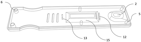
[0084] Such as figure 1 and figure 2 As shown, the collection part includes a casing, a groove 1 arranged on the outer surface of the casing for collecting ocular surface fluid, and a suction hole 2 arranged at the bottom of the groove.

[0085] The groove 1 is arranged at one end of the casing, and the groove 1 forms an opening with the edge of the casing.

[0086] The groove 1 is also provided with a drainage groove 3 , one end of the drainage groove 3 is connected to the edge of the sample suction hole 2 , and the other end extends toward the opening of the groove 1 .

[0087] The outermost edge of the groove is also provided with an anti-overflow strip 4 .

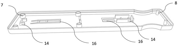
[0088] Such as image 3 and Figure 4 As shown, the housing includes a card cover and a card bottom:

[0089] Both the inner surface of the card cover and the inner surface...

Embodiment 2

[0104] This embodiment provides the method for detecting the specific IgE antibody in the ocular surface fluid sample to be tested by using the colloidal gold qualitative method in embodiment 1:

[0105] 1. Reaction principle

[0106] Anti-IgE monoclonal antibody No. 1 (T line) and goat anti-chicken polyclonal antibody (C line) are pre-immobilized on the nitrocellulose membrane of the immunochromatographic detection card, and anti-IgE monoclonal antibody 2 pre-coated with colloidal gold on the gold standard pad No. and Chicken IgY. After the collected sample of the ocular surface fluid to be tested enters the suction hole 2, the sample of the ocular surface fluid to be tested flows through the sample pad, the gold standard pad and the nitrocellulose membrane according to capillary action. When the sample contains IgE, the target object first forms a reaction complex with the colloidal gold-labeled anti-IgE monoclonal antibody No. 2, and forms a double-antibody sandwich comple...

Embodiment 3

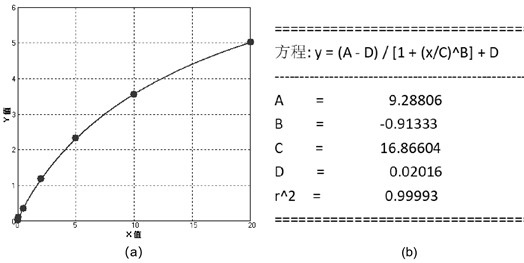
[0125] This embodiment provides the method of detecting the specific IgE antibody in the ocular surface fluid sample to be tested by using the fluorescence quantitative method in embodiment 1:

[0126] 1. Reaction principle

[0127] Anti-IgE monoclonal antibody No. 1 (line T) and goat anti-chicken polyclonal antibody (line C) are pre-fixed on the nitrocellulose membrane, anti-IgE monoclonal antibody No. 2 and chicken polyclonal antibody pre-coated with fluorescent microspheres are pre-coated on the fluorescent pad IgY. After the collected ocular surface fluid sample to be tested enters the suction hole, the ocular surface fluid sample to be tested flows through the sample pad, the fluorescent pad and the nitrocellulose membrane according to capillary action. When the sample contains IgE, the target object first forms a reaction complex with the fluorescently labeled anti-IgE monoclonal antibody No. 2, and forms a double antibody sandwich complex with it when passing through t...


PUM


Abstract

The invention provides an ocular surface liquid collecting and detecting device, and relates to the technical field of medical instruments. Through the design of a groove in a shell, ocular surface liquid of a collected person can be collected at the position of an outer eyelid, direct contact with the ocular surface of a patient is not needed during collection of the ocular surface liquid, uncomfortable reaction of eyes is avoided, and operation is simple and convenient. Meanwhile, ocular surface liquid collected and flowing out from the outer eyelid slides into a sample suction hole in the groove under the action of gravity and then enters a matched immunochromatography detection card, so that related protein factors and pathogenic microorganisms at the ocular surface part can be diagnosed immediately and quickly, the influence of repeated freezing and thawing of protein is avoided, the device has the advantages of simple specific operation, short detection time and the like, detection of related items of the ocular surface liquid can be completed within 15 minutes, the result is accurate, and the problem of on-site diagnosis and treatment of related ocular surface diseases is truly solved.

Description

technical field [0001] The invention relates to the technical field of medical devices, in particular to a device for collecting and detecting ocular surface fluid. Background technique [0002] Immunochromatography is the pre-combination of antibodies labeled with tracer particles (colloidal gold, latex microspheres, fluorescent microspheres, etc.) The antigen or antibody pre-coated on the membrane binds to the flowing complex to develop color. This method can directly interpret the results by naked eyes, and the whole operation is simple and convenient, which is suitable for primary diagnosis and large-scale screening. [0003] The quantitative study of specific protein components in tears has been increasingly used to diagnose ocular surface diseases, judge their severity, and be used as an indication for the prevention or treatment of ocular surface diseases in clinical and scientific research. In actual clinical diagnosis, it is difficult for clinicians to judge the p...

Claims


Application Information

Patent Timeline
Patent Type & Authority Applications(China)
IPC IPC(8): G01N33/558G01N33/53A61B10/00
CPCG01N33/558G01N33/5302A61B10/0045A61B2010/0067
Inventor 陶勇陆成慧常海涛
Owner BEIJING CHAOYANG HOSPITAL CAPITAL MEDICAL UNIV
Why Eureka
Try Eureka