Intelligent analysis system for pathological section image under microscope

A pathological section and microscope technology, applied in image analysis, medical images, analysis materials, etc., can solve problems such as inability to add artificial intelligence algorithms, inability to manually correct processing results, poor local mode processing adaptability, etc., to improve the subjective experience of use. , Improve system real-time, high real-time effect

Pending Publication Date: 2022-03-18
NORTHWEST UNIV(CN)
View PDF6 Cites 1 Cited by
  • Summary
  • Abstract
  • Description
  • Claims
  • Application Information

AI Technical Summary

Problems solved by technology

Since most of these smart microscopes that integrate multiple functions are all-in-one devices, they are not only expensive, but also unable to independently add artificial intelligence algorithms developed by others according to the actual diagnosis of the hospital’s condition.
[0007] 2. After the current artificial intelligence algorithm processes the pathological images, doctors can only passively accept the detection results, and cannot manually correct the processing results
Therefore, doctors often have doubts about this black box mechanism that cannot be manually involved
[0008] 3. Due to differences in microscopes and staining agents between hospitals, most smart microscopes have poor diagnostic results under fixed models, poor adaptability to local model processing, and low accuracy of detection results

Method used


Image

  • Intelligent analysis system for pathological section image under microscope
  • Intelligent analysis system for pathological section image under microscope
  • Intelligent analysis system for pathological section image under microscope

Examples


Embodiment Construction
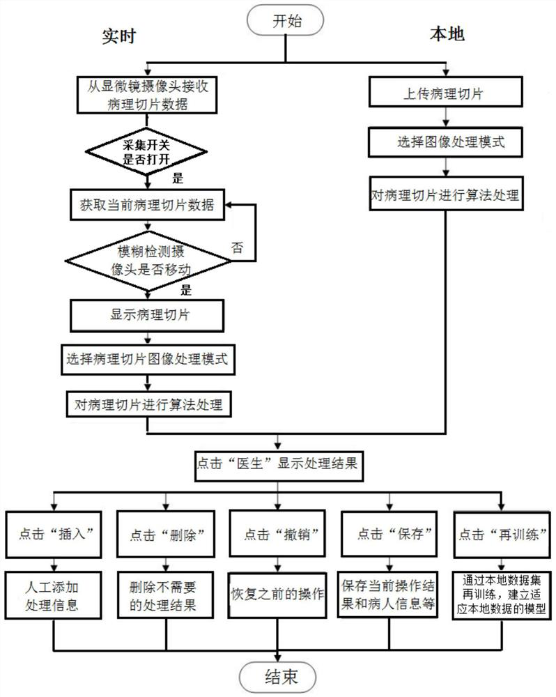
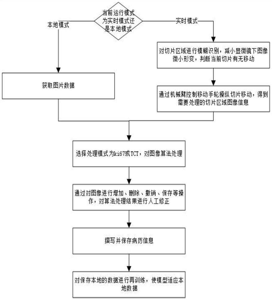
[0072] The design idea of ​​the present invention is to first solve the problem that the existing intelligent microscope cannot remotely move the observation position of the slice, and change the position of the slice by manipulating and moving the hand wheel; immediately after the microscope stops moving, use the fuzzy recognition method to carry out the collected pathological slice image processing, so that the analysis results can be displayed in real time.

[0073] According to the problem that the processing results cannot be manually corrected in the existing problems, and the problem of poor adaptability of the local model is dealt with, an optimization method based on the retraining model is provided to continuously optimize the processing model and improve the accuracy of system analysis.

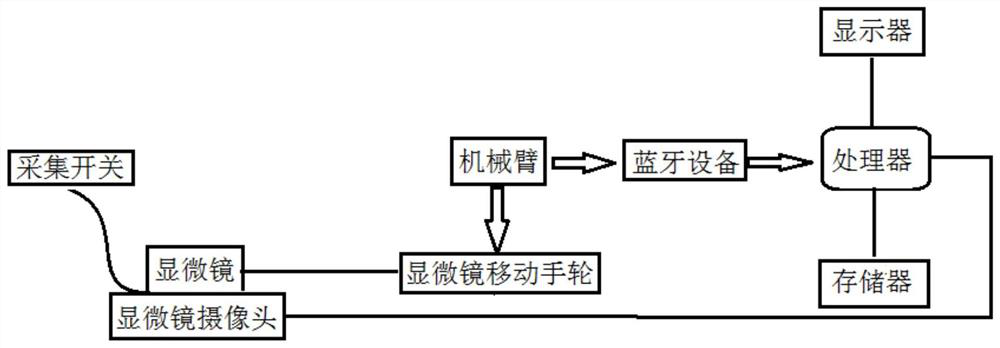
[0074] see figure 1 , the present embodiment provides an intelligent analysis system for pathological slice images under a microscope, including:

[0075] Microscope for observing...


PUM


Abstract

The invention relates to a pathological section image intelligent analysis system under a microscope. The system comprises the microscope, a microscope camera, a mechanical arm, a processor, a memory and a display, the microscope camera is used for collecting images of pathological sections under the microscope, and the mechanical arm achieves section movement under the microscope objective lens. The processor receives a pathological section image from the microscope camera, the system matches an adaptive algorithm model for the digital pathological section image, performs algorithm processing on the pathological section image through intelligent diagnosis to obtain a preliminary diagnosis result, and outputs the labeled pathological section image; carrying out manual judgment on the pathological section image with the label, and if a labeling result meets a requirement, not carrying out correction labeling; otherwise, manually correcting the labeled information of the pathological section image, and outputting a manual judgment result; and processing the manually corrected pathological section image, retraining the model through local data, enhancing the adaptability of the model to the local data, and outputting a final pathological report.

Description

technical field [0001] The invention belongs to the technical field of pathological slice diagnosis, in particular to an intelligent analysis system for pathological slice images under a microscope. Background technique [0002] At present, all kinds of domestic tertiary hospitals and second-level comprehensive, integrated traditional Chinese and Western medicine, foot tumors, children and obstetrics and gynecology specialized hospitals can set up pathology departments if qualified. [0003] However, the efficiency, accuracy, ease of use, and cost-effectiveness of traditional microscopes need to be improved. Doctors need to switch between the screen and the microscope repeatedly, and the judgment results vary from person to person, and pathological sections also rely on doctors to manually identify them. If the field of view under the microscope is digitized, the accuracy and real-time performance can meet the practical application requirements of pathological diagnosis, and...

Claims


Application Information

Patent Timeline
Patent Type & Authority Applications(China)
IPC IPC(8): G06T7/00G06V20/69G06V10/96G06V10/98G06V10/22G06V10/774G06V10/776G16H30/40G16H15/00G16H40/67G01N21/84G01N21/01G06K9/62
CPCG06T7/0012G16H30/40G16H15/00G16H40/67G01N21/84G01N21/01G06T2207/30024G06T2207/30096G06T2207/30168G06F18/2178G06F18/214
Inventor 崔磊刘建业李涵生董阳王亚栋杨林
Owner NORTHWEST UNIV(CN)
Why Eureka
Try Eureka