Medical Imaging Method and System

a medical imaging and image analysis technology, applied in the field of medical imaging methods and systems, can solve the problems of not giving appropriate high “r” values in the rgb system, and the color system of the rgb does not allow expression or normalization of this problem

Active Publication Date: 2008-10-23
YISSUM RES DEV CO OF THE HEBREWUNIVERSITY OF JERUSALEM LTD
View PDF24 Cites 94 Cited by
  • Summary
  • Abstract
  • Description
  • Claims
  • Application Information

AI Technical Summary

Benefits of technology

[0018]It is an object of the present invention to provide a system and method for analysis of changes in color occurring in images of the human body. The inv...

Problems solved by technology

The RGB color system does not allow expression or normalization of this problem.
Images that are truly more intense...

Method used


Image

  • Medical Imaging Method and System
  • Medical Imaging Method and System
  • Medical Imaging Method and System

Examples


examples

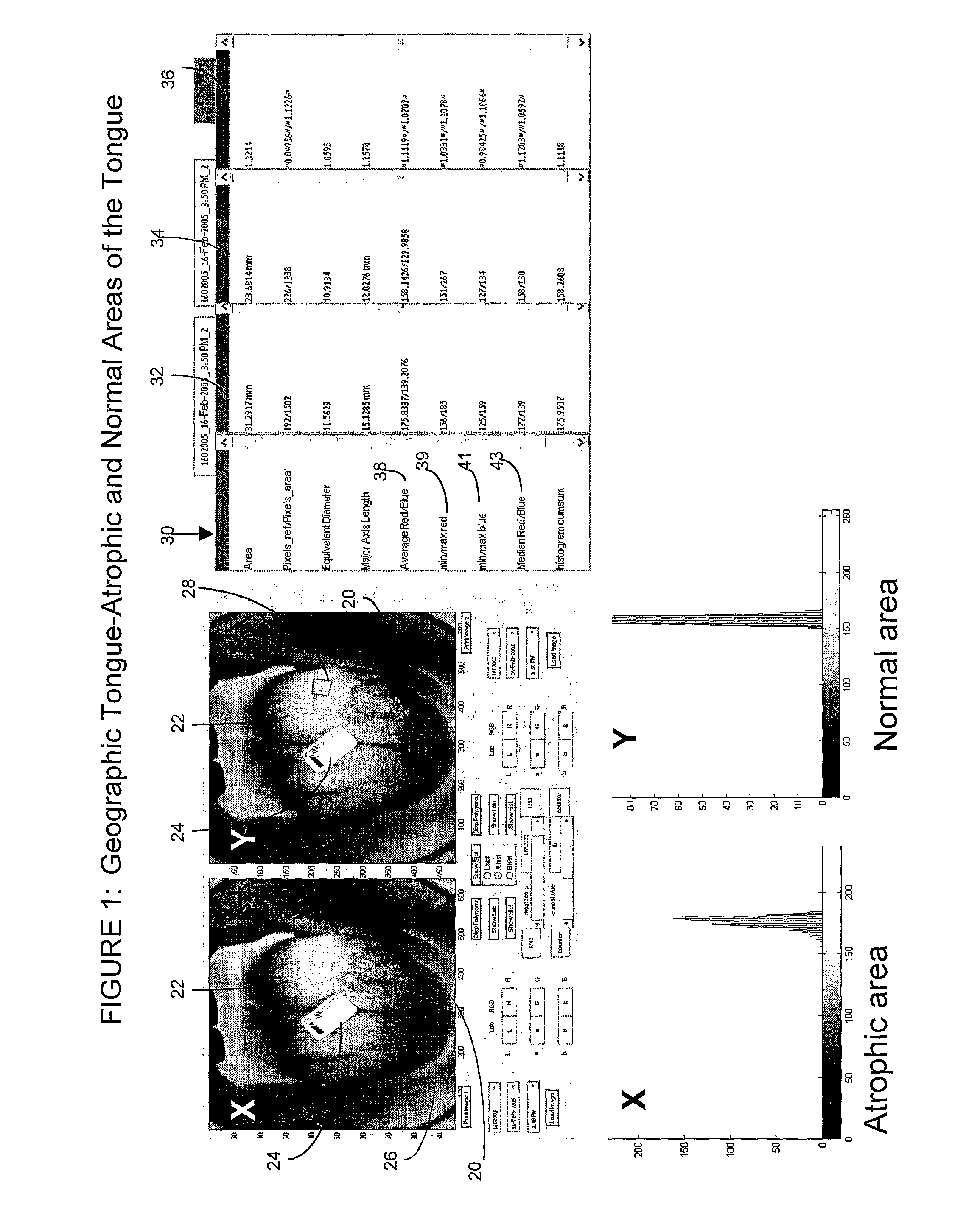
[0140]Referring to FIG. 4, a digital image was captured of a white dysplastic (premalignant) lesion. Note reference label 24 having reference colors, placed adjacent to the lesion 64 before the image was captured. In the left-most panel (referenced “X”), the area of interest selected comprises the lesion. The borders of the lesion have been traced onscreen, and appear with a faint blue outline 66. In the panel referenced “Y”, the digital image is identical, however the area of interest selected comprises normal tissue (faint blue outline 68 above the lesion). Color analysis thus allows comparison between the normal tissue and the lesion.

[0141]Note histogram 70, 72 pertaining to each panel, appearing at bottom left. The color information attributes calculated appear in panel 74 at right, with the results pertaining to the lesion in column 76 (second from the left), and the results pertaining to normal tissue in column 78 (second from the right). A numerical comparison of the lesion a...


PUM


Abstract

A computerized method and system for detection and quantification of changes in color on skin or internal tissue or organs within the human body. Digital images of the skin or tissue containing an area of interest are analyzed using the software of the invention, based on at least one channel of the LAB color system. The invention calculates the intensity and distribution of color present within the predetermined area of interest, and results in display of at least one color information attribute. The invention allows color calibration of the image to be performed in relation to a reference label having predetermined colors which surrounds the area of interest, prior to the analysis. The invention allows a threshold analysis to be performed, which graphically depicts the size and location of areas exhibiting significant change in color, which is a determinant factor in medical diagnosis.

Description

FIELD OF THE INVENTION[0001]The invention relates to image analysis for medical purposes. More specifically, the invention relates to detection and analysis of changes in color on skin or internal tissue or organs within the human body.BACKGROUND OF THE INVENTION[0002]In current practice of dermal analysis and of oral and dental lesion analysis, the physician typically observes the suspected area with the naked eye, or more rarely with an episcope to illuminate the skin, and based on parameters of size, color and other aspects of the visual appearance, decides whether the lesion warrants further observation, medical tests or treatment. The examination and diagnosis are therefore highly subjective, and are dependent upon the medical practitioner's powers of observation and experience.[0003]Occasionally the practitioner will recommend follow up observation in order to ascertain that no changes in size or coloration of the lesion have occurred. These changes suggest malignancy or other...

Claims


Application Information

Patent Timeline
IPC IPC(8): G06K9/00
CPCA61B5/0077A61B5/0088A61B5/1034A61B5/411A61B5/444A61B5/445A61B5/4547G06T7/0012G06T7/408G06T2207/10024G06T2207/30088G06T7/90
Inventor SMITH, YOAVCZERNING, RAKELET
Owner YISSUM RES DEV CO OF THE HEBREWUNIVERSITY OF JERUSALEM LTD
Why Eureka
Try Eureka