

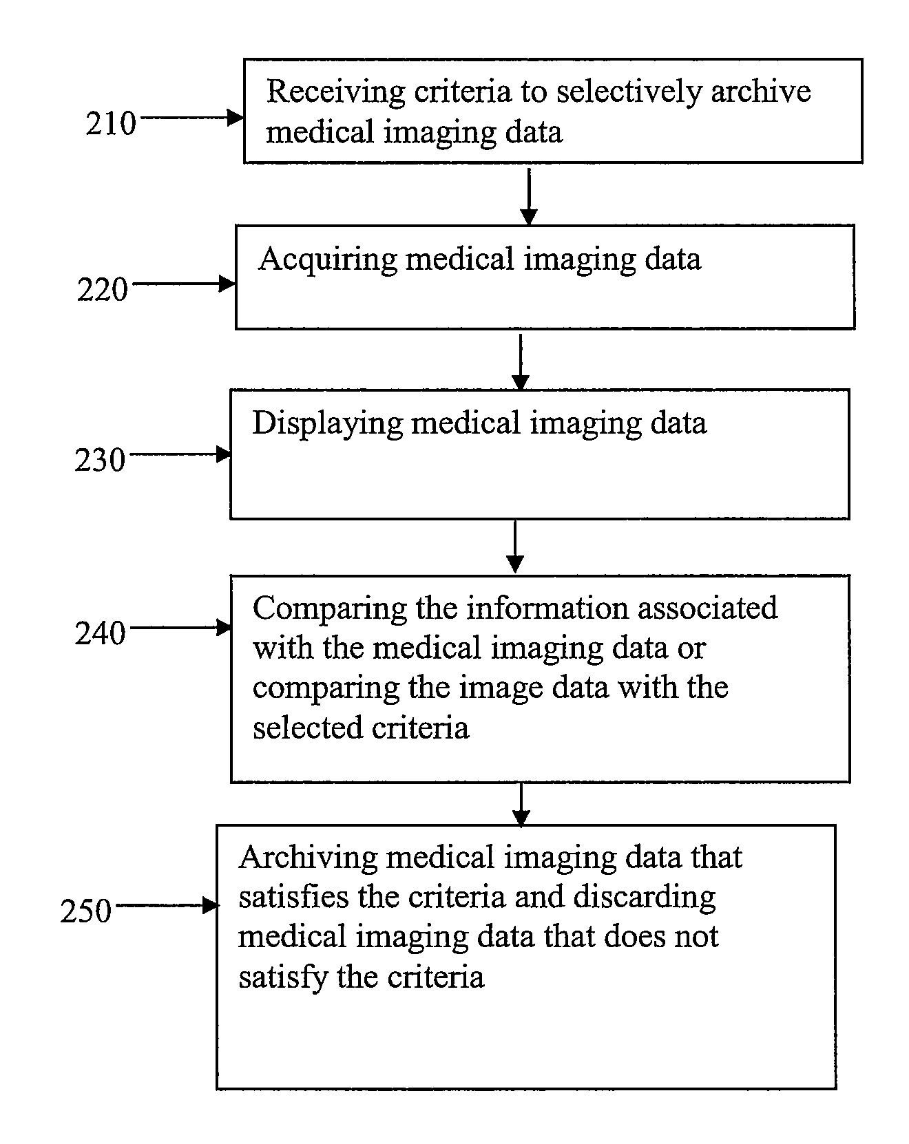
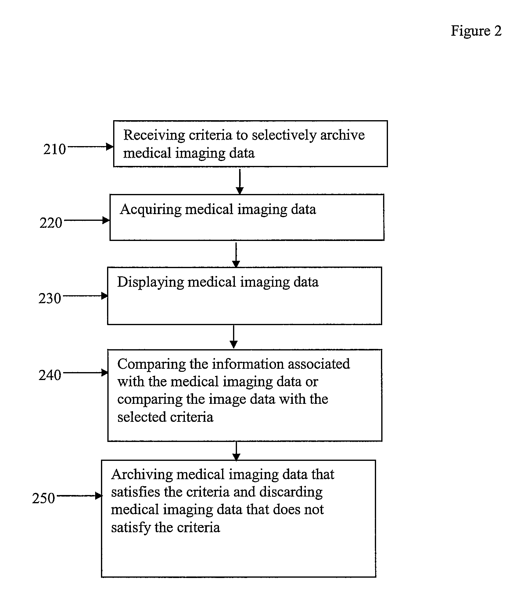
Method and apparatus for flexible archiving
a flexible and archiving technology, applied in the field of system and method for archiving radiology exams, can solve the problems of not being desirable to archive all acquired data, not being desirable to manually select which data, and not being able to detect internal defects in objects
- Summary
- Abstract
- Description
- Claims
- Application Information
AI Technical Summary
Problems solved by technology
Method used
Image

Examples
Embodiment Construction
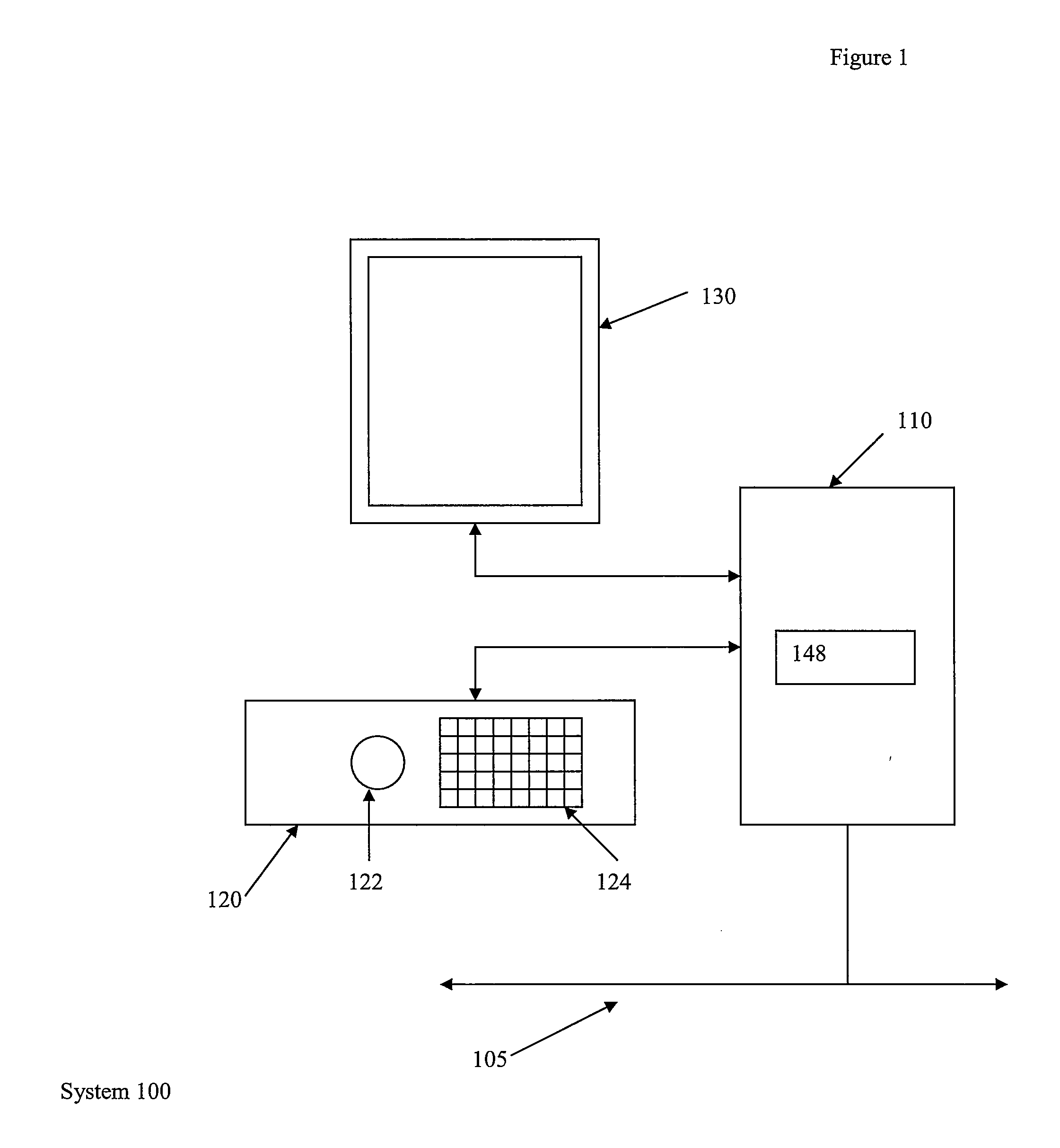
[0009]FIG. 1 illustrates a system 100 for reviewing medical images. The system 100 includes a computer unit 110. The computer unit 110 may be any equipment or software that permits electronic medical images, such as x-rays, ultrasound, CT, MRI, gated MRI, EBT, MR, or nuclear medicine for example, to be electronically acquired, stored, or transmitted for viewing and operation. The computer unit 110 may receive input from a user. The computer unit 110 may be connected to other devices as part of an electronic network. In FIG. 1, the connection to the network is represented by line 105. The computer unit 110 may be connected to network 105 physically, by a wire, or through a wireless medium. In an embodiment, the computer unit 110 may be, or may be part of, a picture archival communication system (PACS).
[0010]The system 100 also includes an input unit 120. The input unit 120 may be a console having a track ball 122 and keyboard 124. Other input devices may be used to receive input from...
PUM
 Abstract
Description
Claims
Application Information