System and method of providing real-time dynamic imagery of a medical procedure site using multiple modalities

a technology of real-time dynamic imagery and medical procedure, applied in the field of system and method of providing real-time dynamic imagery of medical procedure site using multiple modalities, can solve the problems of spatial coordination problem, difficult to perform surgical procedures using mis, and give rise, so as to achieve effective implementation of medical procedures, without undue latency

Active Publication Date: 2010-06-01
INNEROPTIC TECH
View PDF141 Cites 153 Cited by
  • Summary
  • Abstract
  • Description
  • Claims
  • Application Information

AI Technical Summary

Benefits of technology

[0009]The present invention is directed to a system and method of providing composite real-time dynamic imagery of a medical procedure site from multiple modalities which continuously and immediately depicts the current state and condition of the medical procedure site synchronously with respect to each modality and without undue latency. The composite real-time dynamic imagery may be provided by spatially registering multiple real-time dynamic video streams from the multiple modalities to each other. Spatially registering the multiple real-time dynamic video streams to each other may provide a continuous and immediate depiction of the medical procedure site with an unobstructed and detailed view of a region of interest at the medical procedure site. As such, a surgeon, or other medical practitioner, may view a single, accurate, and current composite real-time dynamic imagery of a region of interest at the medical procedure site as he / she performs a medical procedure, and thereby, may properly and effectively implement the medical procedure.
[0010]In this regard, a first real-time dynamic video stream of a scene based on a first modality may be received. A second real-time dynamic video stream of the scene based on a second modality may also be received. The scene may comprise tissues, bones, instruments, and / or other surfaces or objects at a medical procedure site and at multiple depths. The first real-time dynamic video stream and the second real-time dynamic video stream may be spatially registered to each other. Spatially registering the first real-time dynamic video stream and the second real-time dynamic video stream to each other may form a composite representation of the scene. A composite real-time dynamic video stream of the scene may be generated from the composite representation. The composite real-time dynamic video stream may provide a continuous and immediate depiction of the medical procedure site with an unobstructed and detailed view at multiple depths of a region of interest at the medical procedure site. The composite real-time dynamic video stream may be sent to a display.
[0011]The first real-time dynamic video stream may depict the scene from a perspective based on a first spatial state of a first video source. Also, the second real-time dynamic video stream may depict the scene from a perspective based on a second spatial state of a second video source. The first spatial state may comprise a displacement and an orientation of the first video source, while the second spatial state may comprise a displacement and an orientation of the second video source. The first spatial state and the second spatial state may be used to synchronously align a frame of the second real-time dynamic video stream depicting a current perspective of the scene with a frame of the first real-time dynamic video stream depicting a current perspective of the scene. In this manner, the displacement and orientation of the first video source and the displacement and orientation of the second video source may be used to accurately depict the displacement and orientation of the surfaces and objects in the scene from both of the current perspectives in the composite representation.

Problems solved by technology

However, because of limited visibility to certain internal organs, some surgical procedures are at present difficult to perform using MIS.
Operating below while seeing a separate image above can give rise to a number of problems.
These include the issue of parallax, a spatial coordination problem, and a lack of depth perception.
Thus, the surgeon bears a higher cognitive load when employing MIS techniques than with conventional open surgery because the surgeon has to work with a less natural hand-instrument-image coordination.
These problems may be exacerbated when the surgeon wishes to employ other modalities to view the procedure.
But employing more than one modality is often prohibitively difficult to use.
This is a particular problem for modalities like ultrasound, wherein anatomical features do not obviously correspond to the same feature in optical (or white-light) video.
The problems may be further exacerbated in that the surgical site is not static but dynamic, continually changing during the surgery.
Even the amount of gas inside the body cavity (used to make space for the surgical instruments) changes during the surgery, and this affects the shape or position of everything within the surgical site.
Therefore, if the views from the modalities are not continuous and immediate, they may not accurately and effectively depict the current state and / or conditions of the surgical site.
While there is current medical imaging technology that superimposes a video stream using one modality on an image dataset from another modality, the image dataset is static and, therefore, not continuous or immediate.
Periodically updating and / or modifying the image dataset may introduce undue latency in the system, which may be unacceptable from a medical procedure standpoint.
The undue latency may cause the image being viewed on the display by the surgeon to be continually obsolete.
Additionally, relying on the positions of the subject, and / or anatomical or other features and / or landmarks to update and / or modify the image being viewed, may cause the images from the different modalities to not only be obsolete but, also, non-synchronous when viewed.
Accordingly, there currently is no medical imaging technology directed to providing composite real-time dynamic imagery from multiple modalities using two or more video streams, wherein each video stream from each modality may provide a real-time view of the medical procedure site to provide a continuous and immediate view of the current state and condition of the medical procedure site.
Also, there currently is no medical imaging technology directed to providing composite imagery from multiple modalities using two or more video streams, wherein each video stream may be dynamic in that each may be synchronized to the other, and not separately to the position of the subject, and / or anatomical or other features and / or landmarks.
As such, there is currently no medical imaging technology that provides composite real-time, dynamic imagery of the medical procedure site from multiple modalities.

Method used


Image

  • System and method of providing real-time dynamic imagery of a medical procedure site using multiple modalities
  • System and method of providing real-time dynamic imagery of a medical procedure site using multiple modalities
  • System and method of providing real-time dynamic imagery of a medical procedure site using multiple modalities

Examples


Embodiment Construction

[0023]The embodiments set forth below represent the necessary information to enable those skilled in the art to practice the invention and illustrate the best mode of practicing the invention. Upon reading the following description in light of the accompanying drawing figures, those skilled in the art will understand the concepts of the invention and will recognize applications of these concepts not particularly addressed herein. It should be understood that these concepts and applications fall within the scope of the disclosure and the accompanying claims.

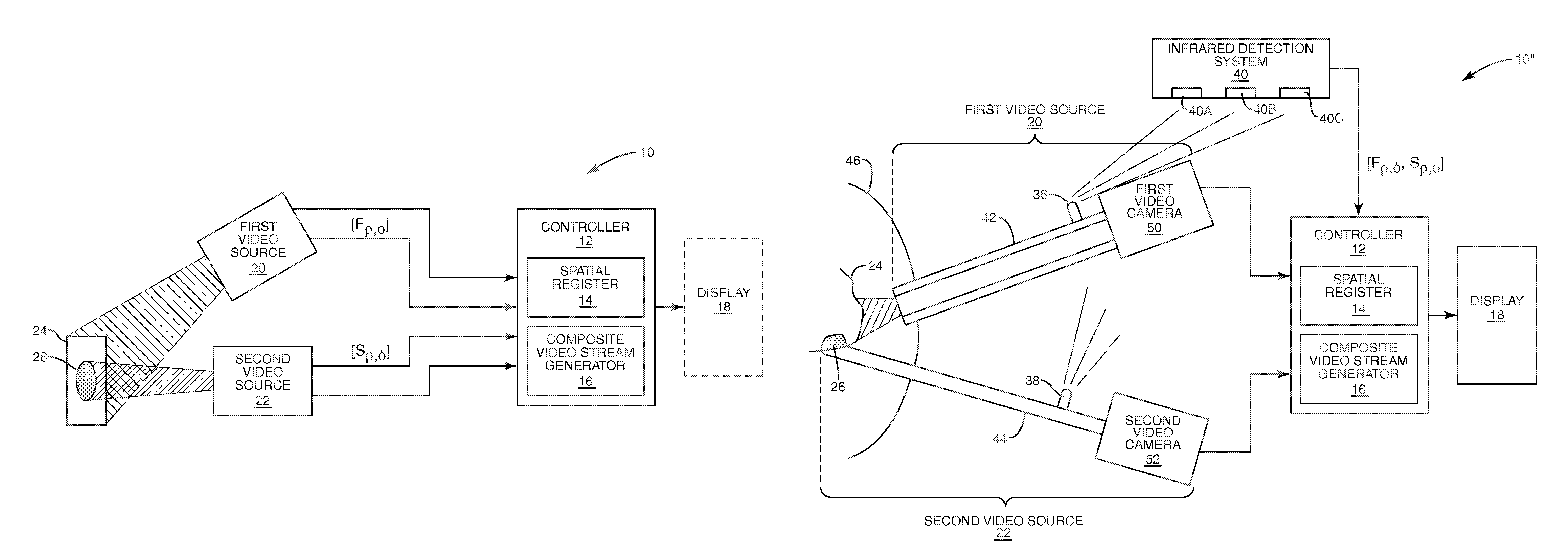
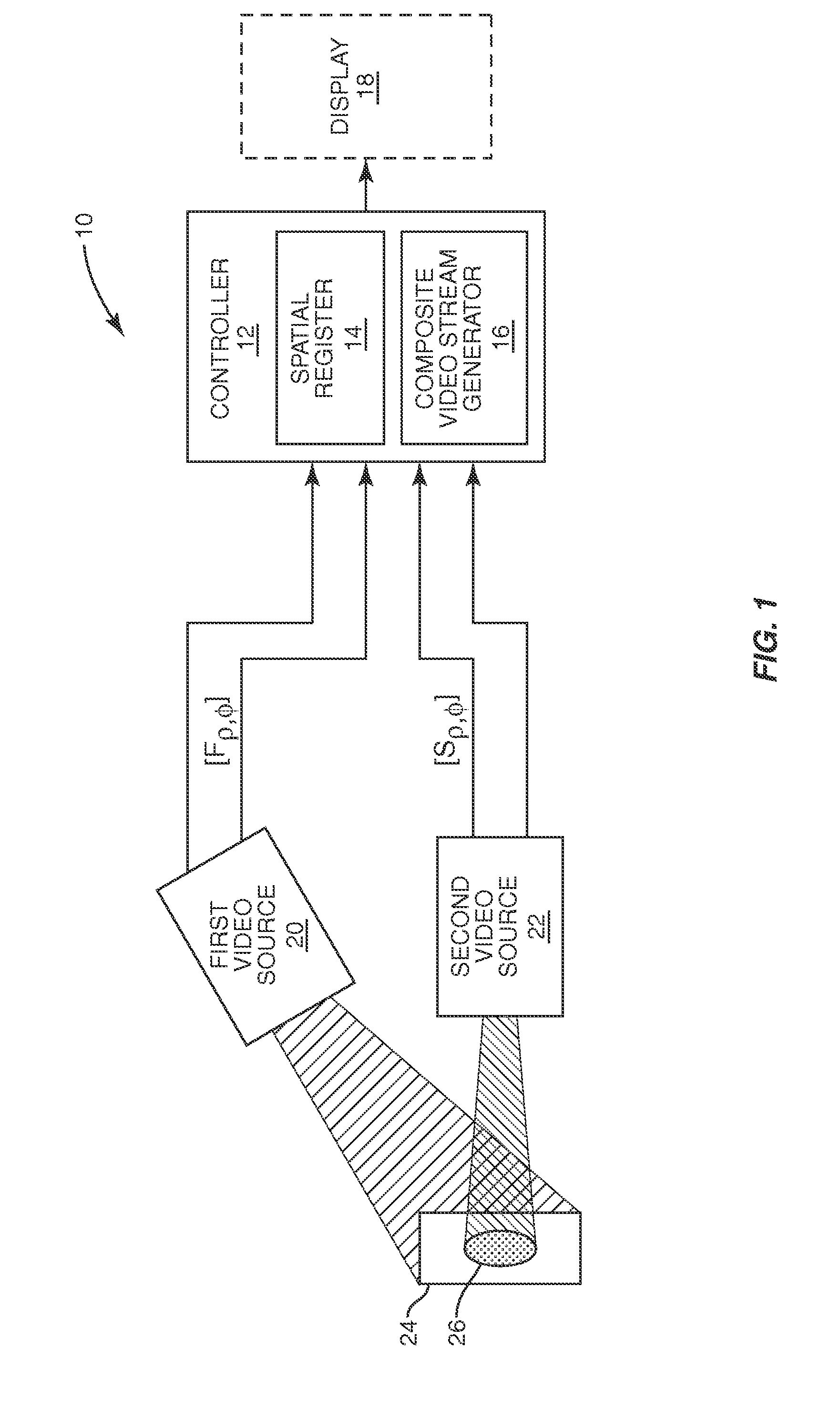
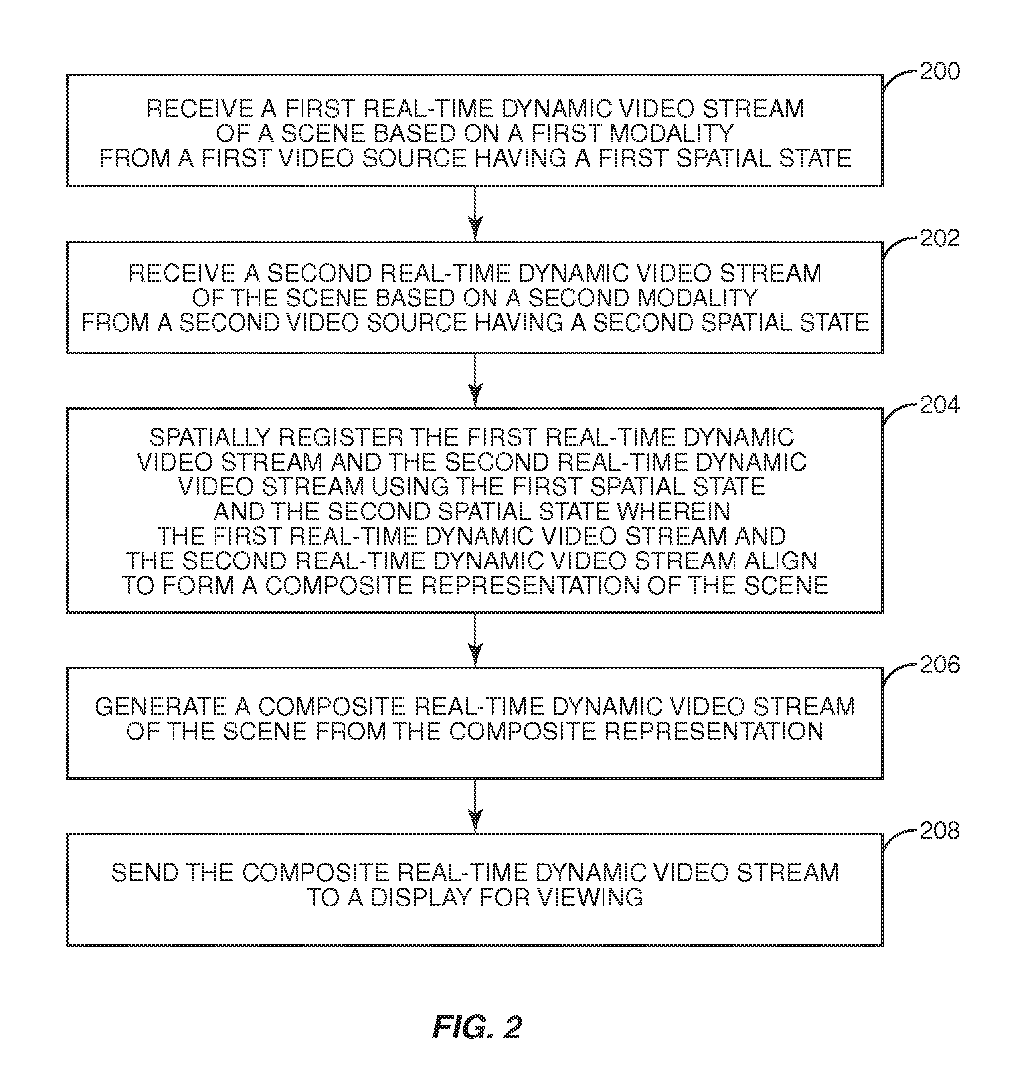
[0024]The present invention is directed to a system and method of providing composite real-time, dynamic imagery of a medical procedure site from multiple modalities which continuously and immediately depicts the current state and condition of the medical procedure site synchronously with respect to each modality and without undue latency. The composite real-time dynamic imagery may be provided by spatially registering multiple re...


PUM


Abstract

A system and method of providing composite real-time dynamic imagery of a medical procedure site from multiple modalities which continuously and immediately depicts the current state and condition of the medical procedure site synchronously with respect to each modality and without undue latency is disclosed. The composite real-time dynamic imagery may be provided by spatially registering multiple real-time dynamic video streams from the multiple modalities to each other. Spatially registering the multiple real-time dynamic video streams to each other may provide a continuous and immediate depiction of the medical procedure site with an unobstructed and detailed view of a region of interest at the medical procedure site at multiple depths. As such, a surgeon, or other medical practitioner, may view a single, accurate, and current composite real-time dynamic imagery of a region of interest at the medical procedure site as he / she performs a medical procedure, and thereby, may properly and effectively implement the medical procedure.

Description

RELATED APPLICATIONS[0001]This application claims the benefit of U.S. Provisional Patent Application Ser. No. 60 / 834,932 entitled “Spatially Registered Ultrasound And Endoscopic Imagery,” filed Aug. 2, 2006, and U.S. Provisional Patent Application Ser. No. 60 / 856,670 entitled “Multiple Depth-Reconstructive Endoscopies Combined With Other Medical Imaging Modalities, And Other Related Technological Details,” filed on Nov. 6, 2006, the disclosures of both of which are hereby incorporated herein by reference in their entireties.FIELD OF THE INVENTION[0002]The present invention is directed to a system and method of providing composite real-time dynamic imagery of a medical procedure site using multiple modalities. One or more of the modalities may provide two-dimensional or three-dimensional imagery.BACKGROUND OF THE INVENTION[0003]It is well established that minimally-invasive surgery (MIS) techniques offer significant health benefits over their analogous laparotomic (or “open”) counter...

Claims


Application Information

Patent Timeline
Patent Type & Authority Patents(United States)
IPC IPC(8): H04N7/18
CPCA61B1/0005A61B1/042G06T3/0087A61B8/463A61B8/5238H04N7/181A61B8/12A61B1/313
Inventor RAZZAQUE, SHARIF A.KELLER, KURTIS P.STATE, ANDREIGREEN, CAROLINE K.ACKERMAN, JEREMY D.
Owner INNEROPTIC TECH
Why Eureka
Try Eureka