Method and apparatus for analyzing elastography of tissue using ultrasound waves

a tissue and ultrasound wave technology, applied in the field of tissue elastography analysis methods and apparatuses, can solve the problem of large measurement error potential

Active Publication Date: 2014-02-13
SAMSUNG ELECTRONICS CO LTD
View PDF8 Cites 10 Cited by
  • Summary
  • Abstract
  • Description
  • Claims
  • Application Information

AI Technical Summary

Problems solved by technology

However, since ultrasound images are read by a medical practitioner with the naked eye, the same ultrasound image may be analyzed differently depending on the angle of view of the medical practitioner, thereby making the potential for a measurement error large.
In addition, occasionally, a medical practitioner incorrectly recognizes abnormal tissue, such as tumorous or cancerous tissue in ultrasound images as normal tissue, that is tissue without tumors or cancer.

Method used


Image

  • Method and apparatus for analyzing elastography of tissue using ultrasound waves
  • Method and apparatus for analyzing elastography of tissue using ultrasound waves
  • Method and apparatus for analyzing elastography of tissue using ultrasound waves

Examples


Embodiment Construction

[0023]Reference will now be made in detail to embodiments, examples of which are illustrated in the accompanying drawings, wherein like reference numerals refer to like elements throughout. In this regard, the present embodiments may have different forms and should not be construed as being limited to the descriptions set forth herein. Accordingly, the embodiments are merely described below, by referring to the figures, to explain aspects of the present description.

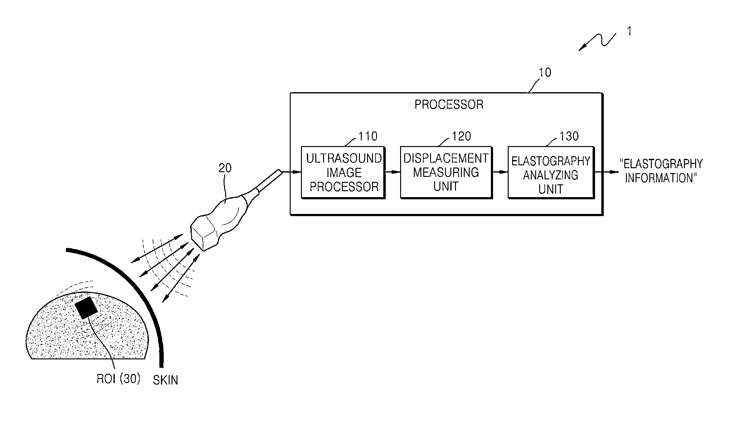
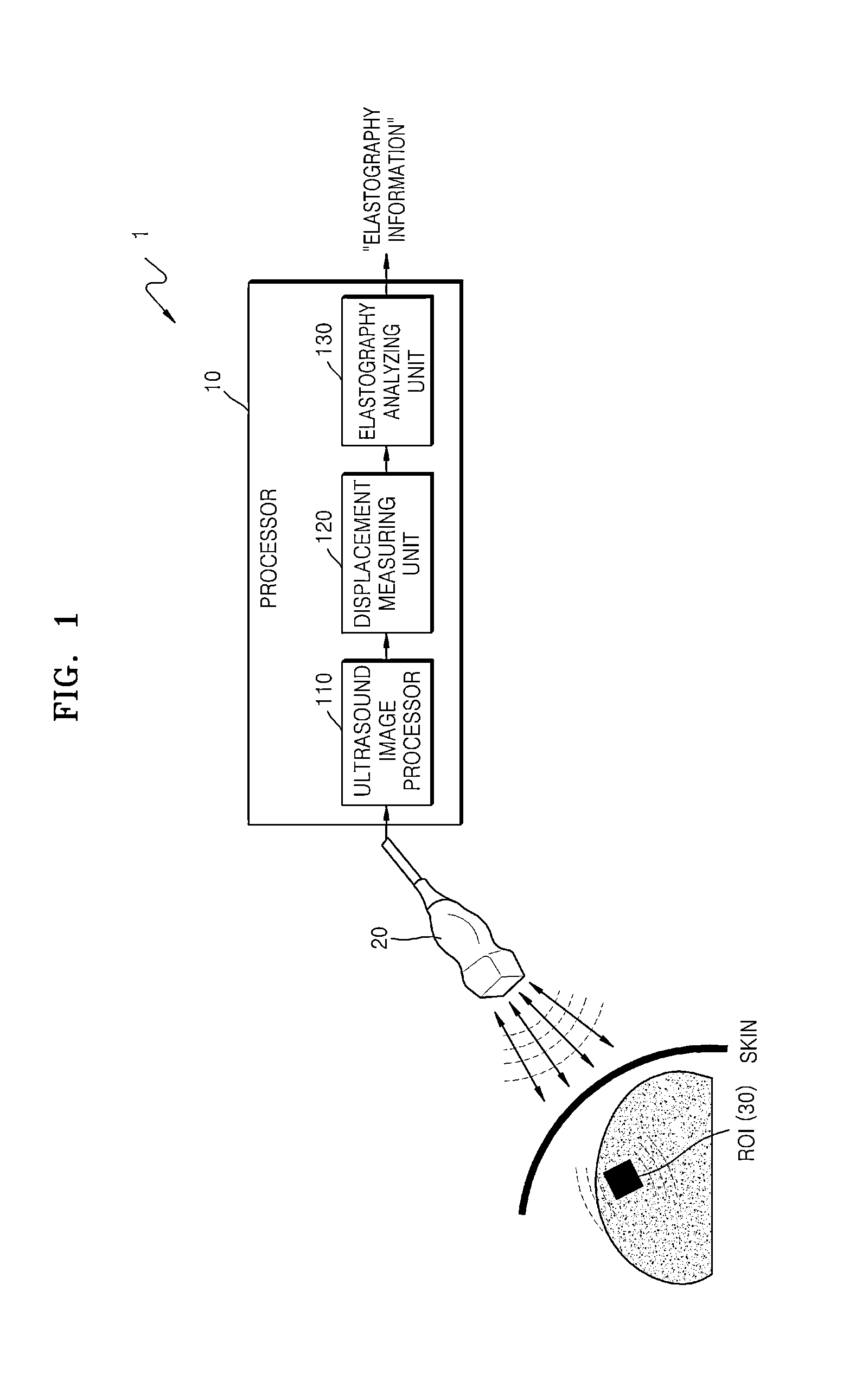
[0024]FIG. 1 is a block diagram of an apparatus 1 for analyzing elastography of tissue using ultrasound waves, according to an embodiment of the present disclosure. Referring to FIG. 1, the apparatus 1 may include, for example, a processor 10 and an ultrasound probe 20. The processor 10 may include, for example, an ultrasound image processor 110, a displacement measuring unit 120, and an elastography analyzing unit 130.

[0025]Only hardware components associated with the current embodiment are described in FIG. 1 to prevent...


PUM


Abstract

A method and apparatus for analyzing elastography of tissue using ultrasound waves, wherein elastography information of tissue in a region of interest (ROI) is analyzed by irradiating ultrasound waves for diagnosis towards the ROI to which a shear wave is induced from an ultrasound probe, receiving echo ultrasound waves, and acquiring three-dimensional (3D) ultrasound images with respect to the ROI.

Description

CROSS-REFERENCE TO RELATED APPLICATIONS[0001]This application claims the benefit of Korean Patent Application No. 10-2012-0086937, filed on Aug. 8, 2012, in the Korean Intellectual Property Office, the disclosure of which is incorporated herein in its entirety by reference.BACKGROUND[0002]1. Field[0003]One or more embodiments of the present disclosure relate to methods and apparatuses for analyzing elastography of tissue of a human or animal subject using ultrasound waves.[0004]2. Description of the Related Art[0005]To diagnose a disease, establish a treatment plan, or evaluate a treatment progress using ultrasound images in medical institutions, a medical practitioner reads ultrasound images of a patient, which are displayed on a monitor, to observe states or sequential histological changes of tumorous or cancerous tissue. However, since ultrasound images are read by a medical practitioner with the naked eye, the same ultrasound image may be analyzed differently depending on the an...

Claims


Application Information

Patent Timeline
Patent Type & Authority Applications(United States)
IPC IPC(8): A61B8/08
CPCA61B8/085A61B8/485A61B8/469A61B8/483A61B8/14G06T7/60
Inventor PARK, JI-YOUNGCHOI, KI-WANLEE, HYOUNG-KIWON, JONG-HWA
Owner SAMSUNG ELECTRONICS CO LTD
Why Eureka
Try Eureka