Method and apparatus for soft-tissue volume visualization

Inactive Publication Date: 2005-05-24
GE MEDICAL SYST GLOBAL TECH CO LLC
View PDF18 Cites 128 Cited by
  • Summary
  • Abstract
  • Description
  • Claims
  • Application Information

AI Technical Summary

Benefits of technology

[0007]In another aspect, a multi-energy computed tomography (MECT) system is provided. The MECT includes at least one radiation source, at least one radiation detector, and a computer operationally coupled to the radiation source and the radiation detector. The computer is configured to receive data regarding a first energy spectrum of a scan of an object, receive data regarding a second energy spectrum of the scan of the object, decompose the received data to generate a first density image representative of bone material and a second density image representative of soft-tissue, identify within the first density image areas smaller than a predetermined size, and import data into the second density image from the data regarding the first energy spectrum according to the identified areas of the first density image.
[0008]In a further aspect, a multi-energy computed tomography (MECT) system is provided. The CT system includes at least one radiation source, at least one radiation detector, and a computer operationally coupled to the radiation source and the radiation detector. The computer is configured to receive image data for an object, decompose the received image data into a first

Problems solved by technology

In spite of recent advancements in computed tomography (CT) technology, such as faster scanning speed, larger coverage with multiple detector rows, and thinner slices, energy resolution is still a missing piece, namely, a wide x-ray photon energy spectrum from the x-ray source and a lack of energy resolution from CT detection systems preclude energy discrimination CT.
Some beam-hardening artifacts can be easily corrected, but others may be more difficult to correct.
However, beam

Method used


Image

  • Method and apparatus for soft-tissue volume visualization
  • Method and apparatus for soft-tissue volume visualization
  • Method and apparatus for soft-tissue volume visualization

Examples


Example

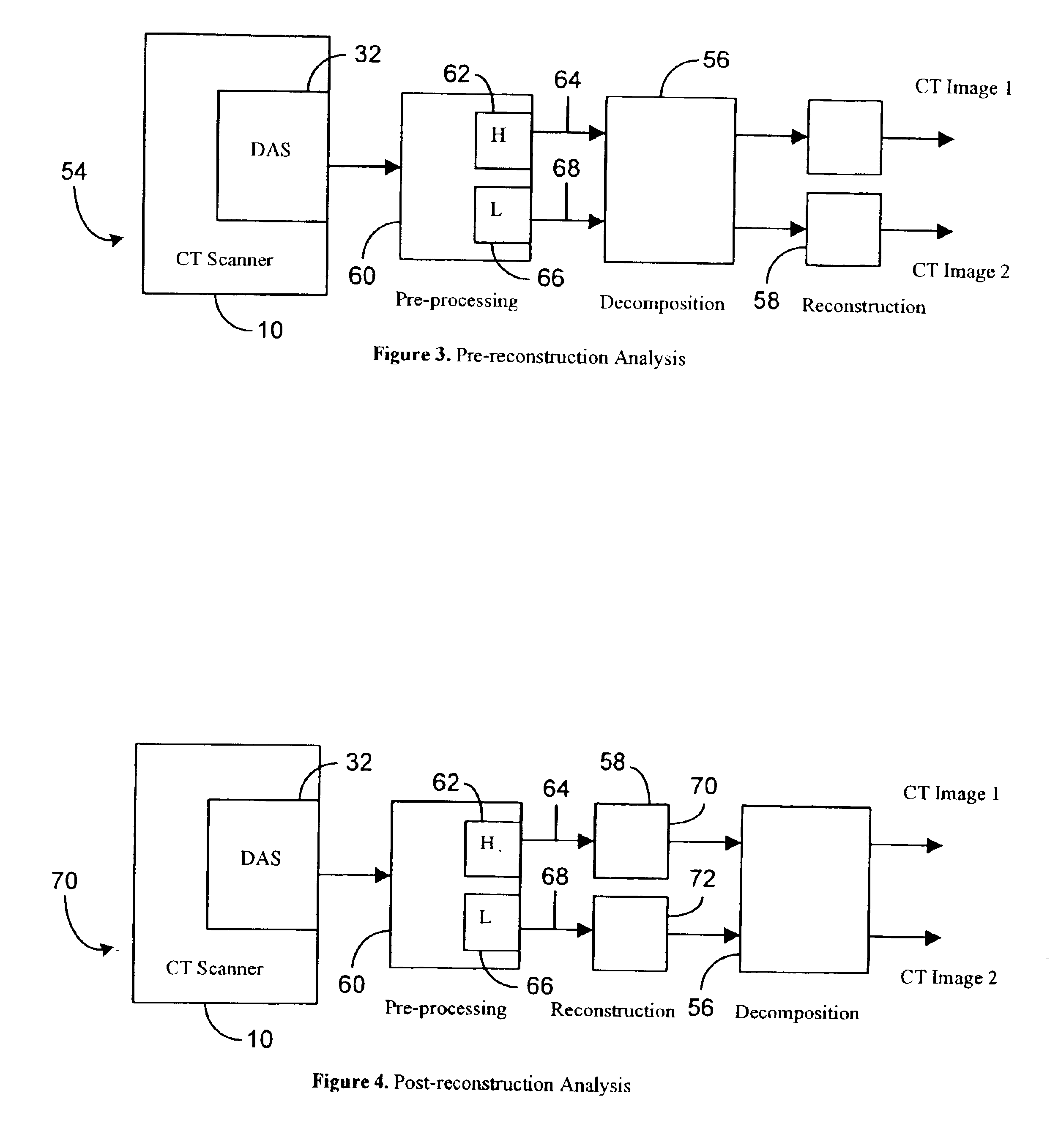
[0021]The methods and apparatus described herein facilitate augmenting segmentation capabilities of multi-energy imaging with a method for image-based segmentation. The methods and systems described herein facilitate real-time volume buildup and visualization of soft-tissue. More specifically, the methods and systems described herein facilitate segmenting bone material from an image while retaining calcification within the image, and facilitate augmenting segmentation capabilities of multi-energy imaging to guide surgical navigation and radiation therapy.

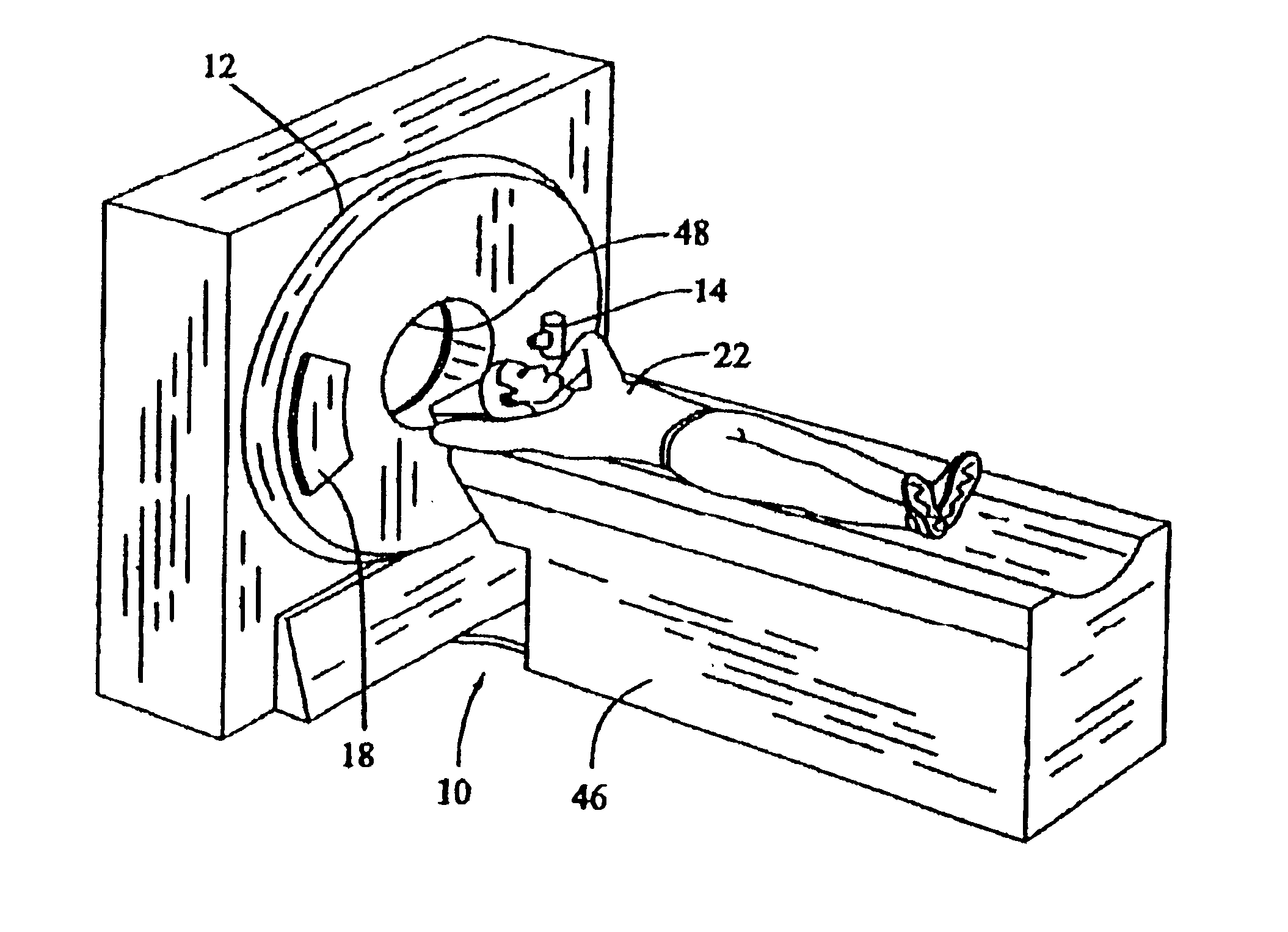
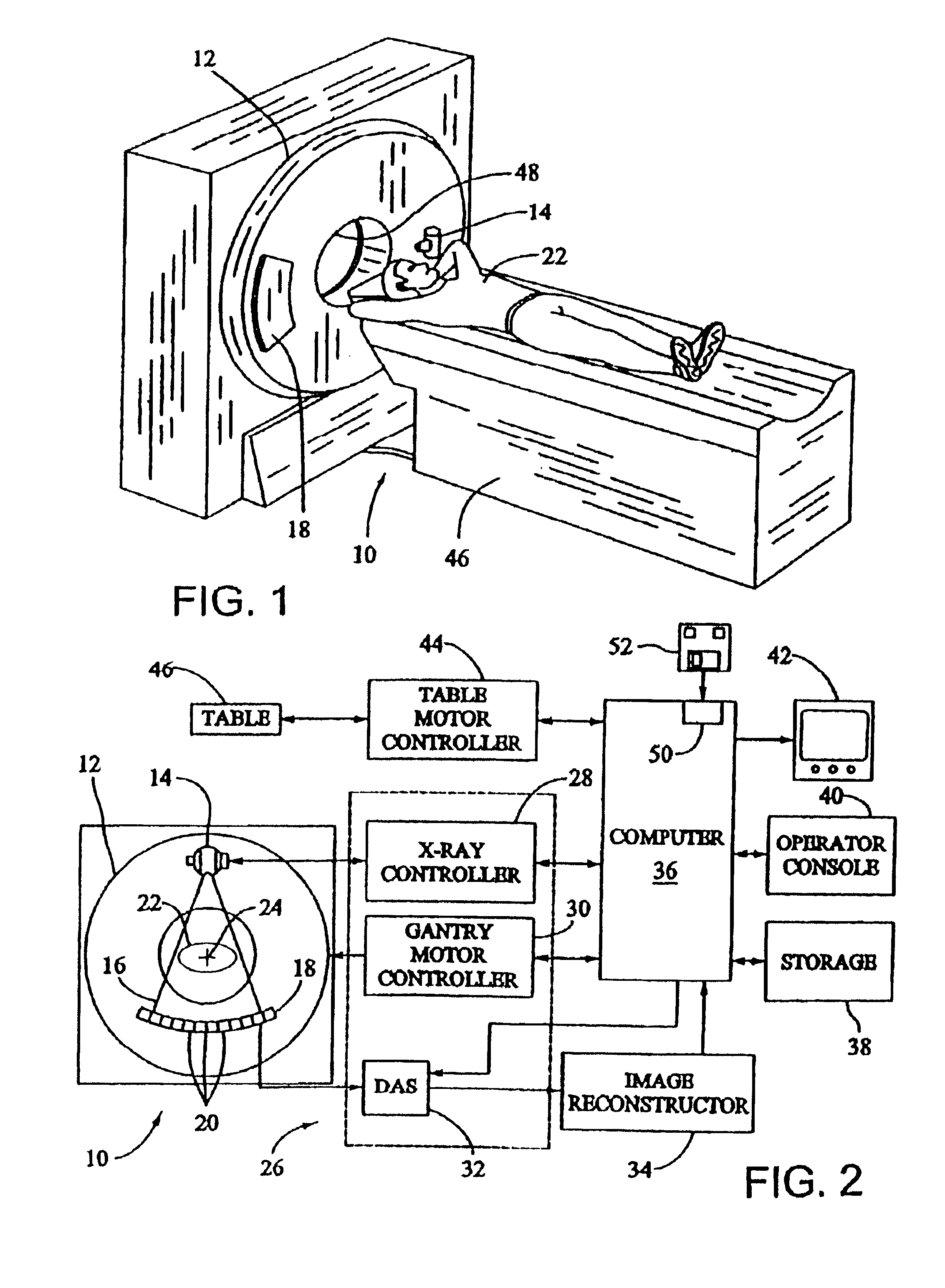
[0022]In some known CT imaging system configurations, an x-ray source projects a fan-shaped beam which is collimated to lie within an x-y plane of a Cartesian coordinate system and generally referred to as an “imaging plane”. The x-ray beam passes through an object being imaged, such as a patient. The beam, after being attenuated by the object, impinges upon an array of radiation detectors. The intensity of the attenuated radiation ...


PUM


Abstract

A method for obtaining data including scanning an object using a multi-energy computed tomography (MECT) system to obtain data to generate an anatomical image, and decomposing the obtained data to generate a first density image representative of bone material and a second density image representative of soft-tissue. The method further includes segmenting at least one of the first density image and the second density image, and volume rendering the second density image.

Description

BACKGROUND OF THE INVENTION[0001]This invention relates generally to medical imaging systems, and more specifically to a method and apparatus for soft-tissue volume visualization using a medical imaging system.[0002]In spite of recent advancements in computed tomography (CT) technology, such as faster scanning speed, larger coverage with multiple detector rows, and thinner slices, energy resolution is still a missing piece, namely, a wide x-ray photon energy spectrum from the x-ray source and a lack of energy resolution from CT detection systems preclude energy discrimination CT.[0003]X-ray attenuation through a given object is not a constant. Rather, x-ray attenuation is strongly dependent on the x-ray photon energy. This physical phenomenon manifests itself in an image as a beam-hardening artifact, such as non-uniformity, shading, and streaks. Some beam-hardening artifacts can be easily corrected, but others may be more difficult to correct. In general, known methods to correct be...

Claims


Application Information

Patent Timeline
IPC IPC(8): A61B6/03A61B6/12G01V5/00
CPCA61B6/032A61B6/4035A61B6/405A61B6/463A61B6/466A61B6/482A61B6/12Y10S378/901A61B6/4241
Inventor AVINASH, GOPAL B.SABOL, JOHN MICHAELWALKER, MATTHEW JOSEPH
Owner GE MEDICAL SYST GLOBAL TECH CO LLC
Why Eureka
Try Eureka