Display for computer-aided diagnosis of mammograms

a mammogram and computer-aided technology, applied in the field of mammogram computer-aided diagnosis, can solve the problems of difficult to see or evaluate lesions, require advanced diagnostic tools, and difficult to interpret mammograms

Inactive Publication Date: 2007-04-10
SIEMENS COMP AIDED DIAGNOSIS
View PDF17 Cites 17 Cited by
  • Summary
  • Abstract
  • Description
  • Claims
  • Application Information

AI Technical Summary

Benefits of technology

[0032]It is an object of the present invention to provide a system and method for displaying data which assist a ra

Problems solved by technology

Since the interpretation of mammographic lesions is problematic, a need for advanced diagnostic tools is required.
The analysis, even after using the above techniques, still remains mainly subjective.
Reading large numbers of mammograms is a difficult and tiring task.
According to some literature reports as noted above, unacceptably high rates of false negative results occur.
However, it is well known that computer-aided detection systems almost invariably indicate more suspected abnormalities than are detected by a trained radiologist.
Therefore, a radiologist may have to examine, and must often dismiss, not only the suspected abnormalities that he detects from the radiological mammog

Method used


Image

  • Display for computer-aided diagnosis of mammograms
  • Display for computer-aided diagnosis of mammograms
  • Display for computer-aided diagnosis of mammograms

Examples


Embodiment Construction

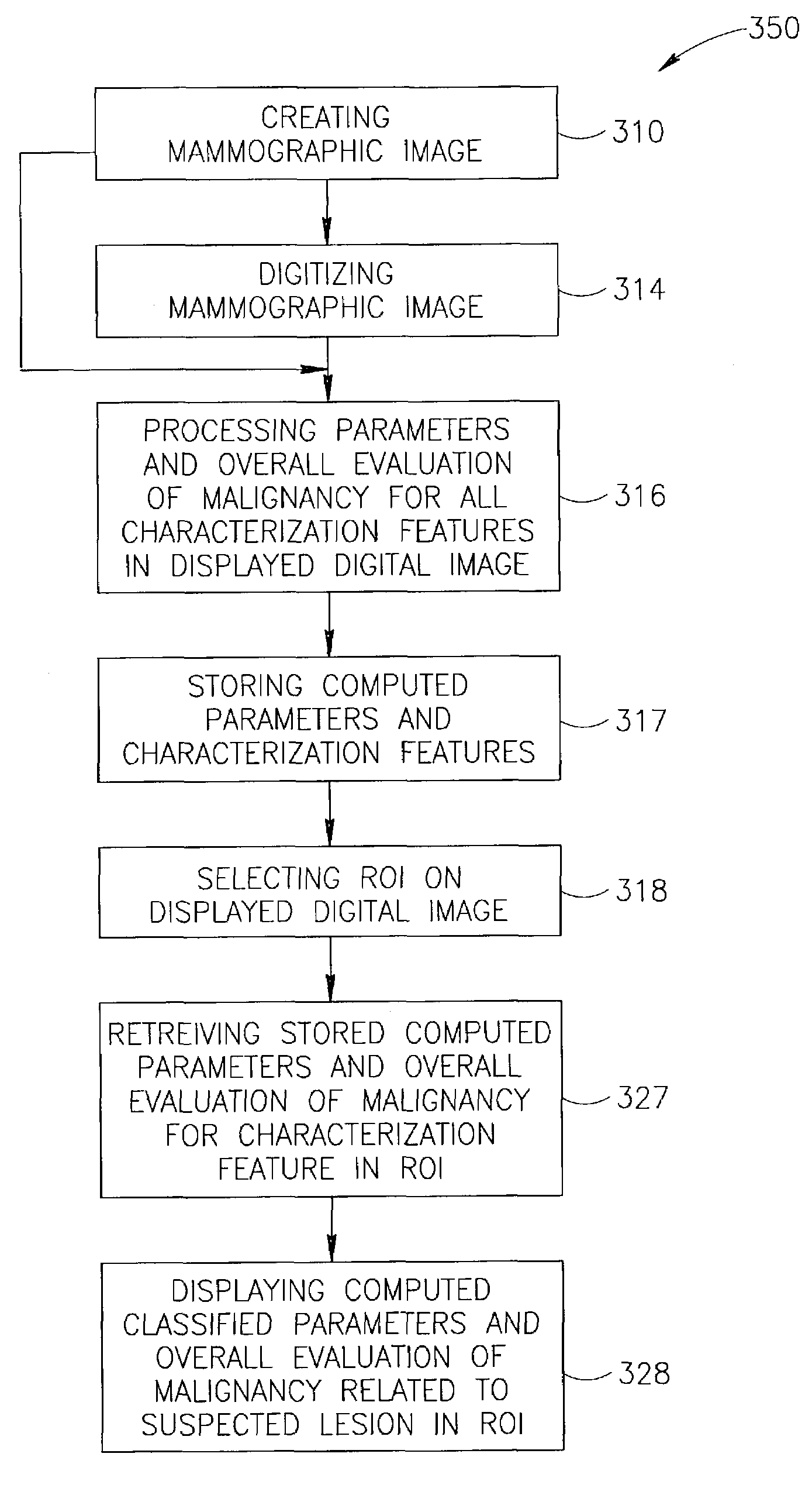
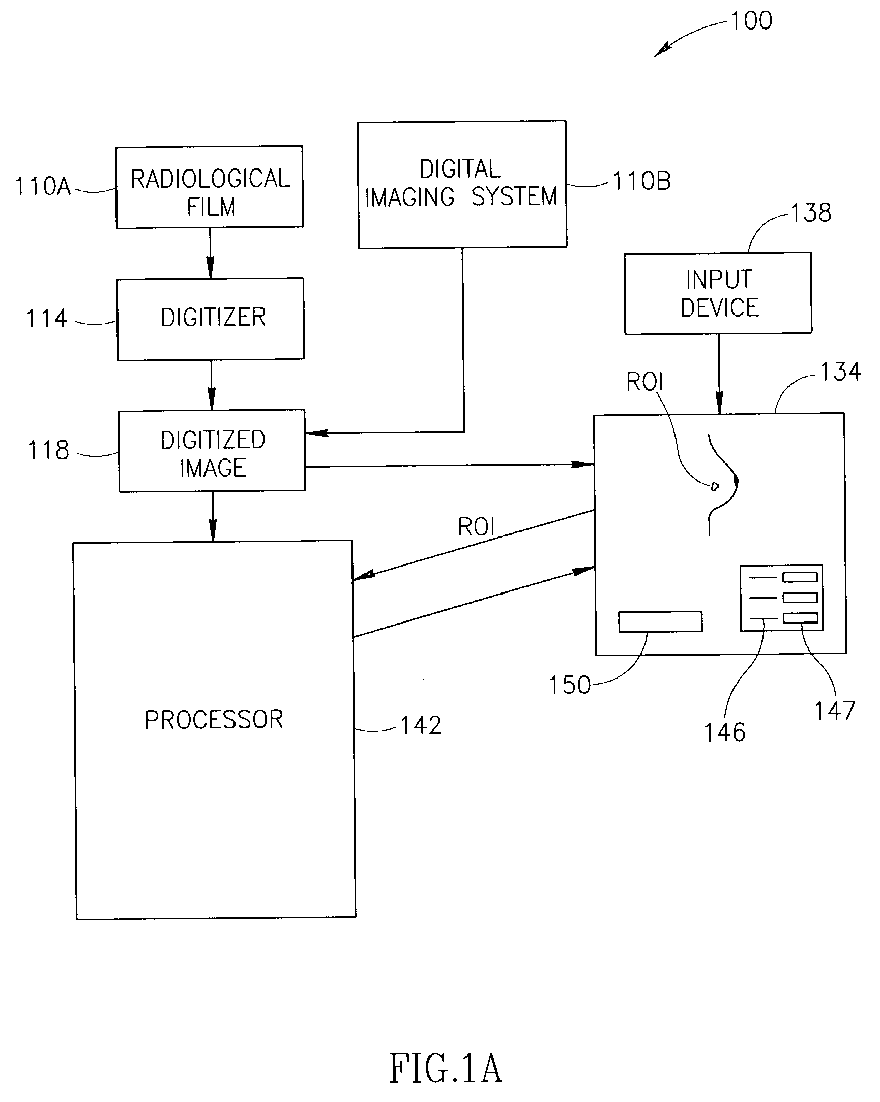
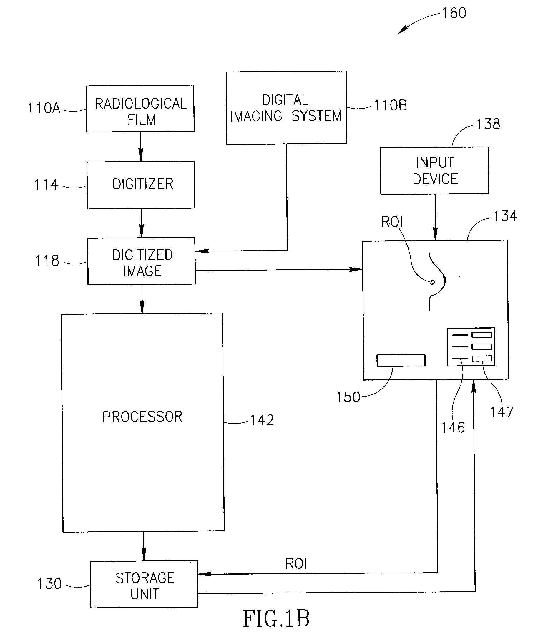
[0048]The present invention relates to a method and system for displaying digitized mammogram images and diagnosis-assisting information that aids in interpreting the images. More specifically, the invention relates to a computer-aided diagnosis (herein after sometimes denoted as “CAD”) method and system for classifying and displaying malignancy evaluation / classification data for anatomical abnormalities in digitized mammogram images. Characterization features of suspected abnormalities in user-selected regions of interest (ROI) are viewed on a display in conjunction with an overall evaluation of malignancy and usually also with a plurality of quantified parameters related to the characterization features. The overall evaluation of malignancy and / or the plurality of quantified parameters are herein also called classifier data. The characterization features viewed and evaluated / classified are also user-selected.

[0049]The overall evaluation of a suspected lesion in the radiological im...


PUM

PropertyMeasurementUnit
Timeaaaaaaaaaa
Densityaaaaaaaaaa

Abstract

A method for displaying a computer-generated determination of the likelihood of malignancy in a mammogram lesion. The method requires providing a digitized image of a mammogram, displaying the digitized image, and selecting a region of interest directly on the displayed digitized image. The digitized image is then processed so that classifier data of the lesion in the user-selected region of interest are generated and displayed. A system for displaying a determination of the likelihood of malignancy in a mammogram lesion. The system includes a display for presenting a digitized mammogram and an input device in communication with the display for selectably indicating a region of interest on the displayed mammogram. The system also includes a processor for generating classifier data related to a characterization feature within the region of interest. The classifier data is presented on the display.

Description

FIELD OF THE INVENTION[0001]The present invention relates to a method and system for displaying evaluation / classification information that assists in determining the malignancy or non-malignancy of abnormalities appearing on radiological mammogram images.BACKGROUND OF THE INVENTION[0002]Breast cancer is one of the most common types of cancer afflicting Western society. It is estimated that the spread of the disease has risen in the United States, from one in twenty women being afflicted in 1940, to one in eight in 1995. The American Cancer Society estimated that 183,000 new cases of breast cancer were reported during 1995. In the United States, some 46,000 women die from the disease per year. Today, it is accepted that the best way to detect breast cancer in its early stages is by annual mammography screening of women aged 40 and up.[0003]The five-year survival rate for localized breast cancer is 93%. That rate drops to 72% if the cancer has spread regionally by the time of diagnosi...

Claims


Application Information

Patent Timeline
IPC IPC(8): G06K9/00A61B5/055A61B6/00A61B10/00G06F17/00G06T7/00
CPCG06K9/033G06T7/0012G06K2209/05G06T2207/30068G06V10/987G06V2201/03
Inventor LEICHTER, ISAACBAMBERGER, PHILIPPENOVAK, BORIS
Owner SIEMENS COMP AIDED DIAGNOSIS
Why Eureka
Try Eureka