Method and apparatus for vasculature visualization with applications in neurosurgery and neurology

a vasculature and neurology technology, applied in the field of medical imaging, can solve the problems of avm enlargement, increased risk of bleeding again in the future, profound disabling or fatal hemorrhage,

Active Publication Date: 2012-05-22
STRYKER EUROPEAN OPERATIONS LIMITED
View PDF49 Cites 91 Cited by
  • Summary
  • Abstract
  • Description
  • Claims
  • Application Information

AI Technical Summary

Problems solved by technology

Over time, the AVM tends to enlarge as the great pressure of the arterial vessels can not be handled by the veins that drain out of it.
When symptoms do occur before an AVM ruptures, they are related to smaller and slower bleeding from the abnormal vessels, which are often fragile because their structure is abnormal.
Depending on the location and the severity of the bleed, the hemorrhage can be profoundly disabling or fatal.
If an AVM bleeds once, the risk is greater that it will bleed again in the future.
Very large AVMs may short-circuit blood flow enough to cause cardiac decompensation in which the heart is unable to pump enough blood to compensate for bleeding in the brain.
Traditional imaging methods used in the context of AVM, such as measuring arterial pulsation or angiography using digital subtraction are either unreliable or expensive and inconvenient to be used during surgery (see, e.g., Wrobel et al., 1994, Neurosurgery 35(5):970; Martin et al., 1990, J. Neurosurg. 73:526).

Method used


Image

  • Method and apparatus for vasculature visualization with applications in neurosurgery and neurology
  • Method and apparatus for vasculature visualization with applications in neurosurgery and neurology
  • Method and apparatus for vasculature visualization with applications in neurosurgery and neurology

Examples


example 1

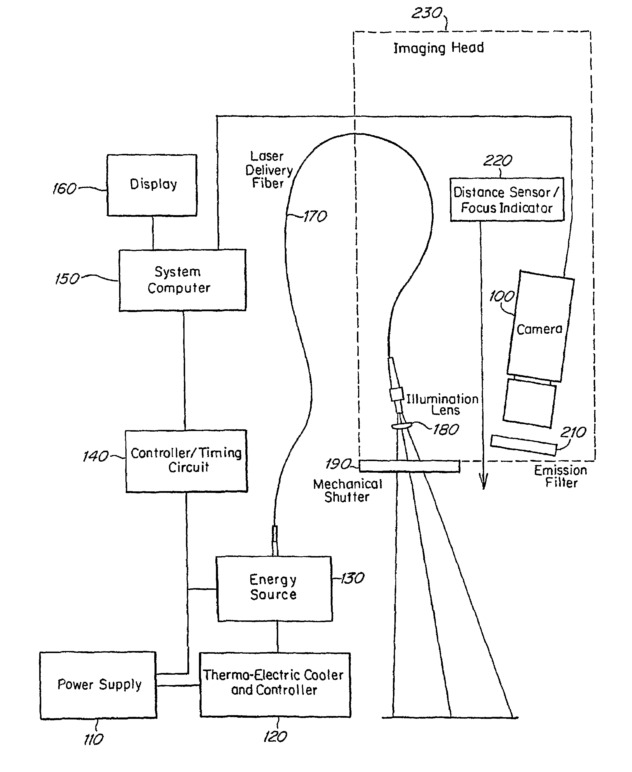
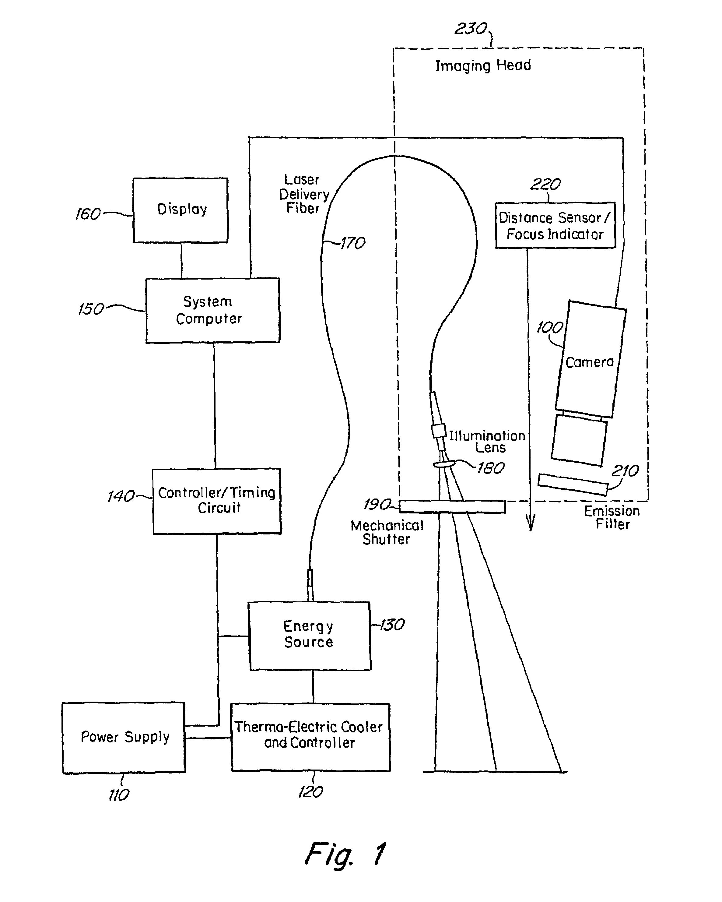
[0086]This example illustrates a system of the invention. The imaging devices is made primarily of two subsystems that are primarily optical in nature, an illumination subsystem and a detection subsystem. Other subsystems are primarily electrical or mechanical in nature. The illumination subsystem includes a fiber-coupled, infrared laser, a light guide, and a projector lens. The detection subsystem includes a high-quality imaging lens, a narrow band-pass filter, and a CCD camera. The remainder of the system includes a laser, video display, computer and other auxiliary control circuits. The system is designed with an articulated arm with an imaging head. The imaging head contains the imaging and illumination optics and electronics. The articulated arm allows the illumination and imaging systems to be positioned over the field of operation.

[0087]Mounted inside this imaging head are the filter and CCD camera, a distance sensor and the light guide and projection lens. A power delivery o...


PUM


Abstract

The invention provides methods and systems for neurovascular imaging (150).

Description

RELATED APPLICATIONS[0001]This application claims the benefit of PCT Application Ser. No. PCT / US2006 / 016101, filed Apr. 26, 2006; which claims the benefit of U.S. provisional Ser. No. 60 / 674,890, filed Apr. 26, 2005, the disclosures of all of which are incorporated herein by reference in their entirety as if fully set forth herein.FIELD OF THE INVENTION[0002]The invention relates generally to the field of medical imaging. Certain embodiments of the invention provide methods for imaging vasculature in a subject. Certain other embodiments provide systems which are useful for imaging vasculature in a subject.BACKGROUND OF THE INVENTION[0003]Imaging of biological tissues and organs assists doctors in both diagnosis and treatment. A variety of medical techniques which are suitable for imaging biological tissues and organs are known. These include traditional x-rays, ultra-sound, as well as magnetic resonance imaging (MRI), and computerized tomography (CT). A variety of dyes used in medic...

Claims


Application Information

Patent Timeline
Patent Type & Authority Patents(United States)
IPC IPC(8): A61B5/00
CPCA61B5/0261A61B5/103G06K2009/00932A61B5/4064G06V40/14
Inventor MANGAT, GURPREETBRZOZOWSKI, LUKASZCROFT, RICHARD L.
Owner STRYKER EUROPEAN OPERATIONS LIMITED
Why Eureka
Try Eureka