Endoscope accessory

a technology for endoscopes and accessories, applied in the field of endoscope accessories, can solve the problems of poor image quality and hammer obtaining ultrasonic images, and achieve the effects of improving the ability of endoscopes to maintain luminal views, improving acoustic coupling, and improving examination

Active Publication Date: 2018-01-16
IZOMED INC
View PDF84 Cites 4 Cited by
  • Summary
  • Abstract
  • Description
  • Claims
  • Application Information

AI Technical Summary

Benefits of technology

[0003]This device presented in this invention enhances capabilities of an endoscope in maintaining luminal view and is comprised of an overtube with an inflatable, freely moveable positioning balloon at its distal endportion. The overtube can slide over an endoscope inside the body cavity, and its distal tip is placed just proximal to the distal tip of endoscope, proximal to the portion of the gastrointestinal tract that needs to be examined. At this place the positioning balloon is inflated to secure the position of the overtube. The other part of the device is a catheter with an occlusion balloon at its distal endportion that can be passed through a passageway provided along the overtube and extends beyond the tip of the endoscope distal to the portion of the gastrointestinal tract that needs to be examined. At this position, the occlusion balloon is inflated. This creates a closed space between the two inflated balloon in the gastrointestinal tract at the portion of the gastrointestinal tract that needs to be examined. This closed space can be inflated with air for detailed examination and treatment of the area with a regular endoscope, or filled with water for acoustic coupling between the endoscope transducer and gastrointestinal structures for examination with an echoendoscope. The inflated balloons prevent escape of air or water from the examination site. This improves the acoustic coupling and provides for better examination in a larger region of the gastrointestinal tract that needs to be examined. This reduces unnecessary overinflation of the other portion of the gastrointestinal tract with air and water, and in the case of water, reduces the risk such as water toxicity or aspiration. Throughout the procedure, the pressure of the water or air in the closed space is monitored to prevent overdistension of the region with water or air. After termination of the examination, the water or air is suctioned out of the part of the gastrointestinal tract examined.

Problems solved by technology

However, air create a barrier to ultrasonic was passage and hampers obtaing ultrasonic images.
In most parts of the gastrointnal tract, however, the large diameter of the lumen and or the angle of the transducer in relation to the intestinal wall result in an inadequate contact between the transducer balloon and the intestinal wall and thus, the operator usually use water infusion to fill the portion of the gastrointestinal tract with water and create acoustic coupling between the transducer and the examinimed structures.
Unfortunately, the gastrointestinal tract is not a closed region and the infused water soon moves to other portion of the gastrointestinal tract and this can often result in poor image quality despite repeated infusion of water around the transducer.
In addition, infusion of significant amount of water during the examination could result in untoward problems such as aspiration of the water into the patient's airway or overdistention of the gastrointestinal tract.

Method used


Image

  • Endoscope accessory
  • Endoscope accessory
  • Endoscope accessory

Examples


Embodiment Construction

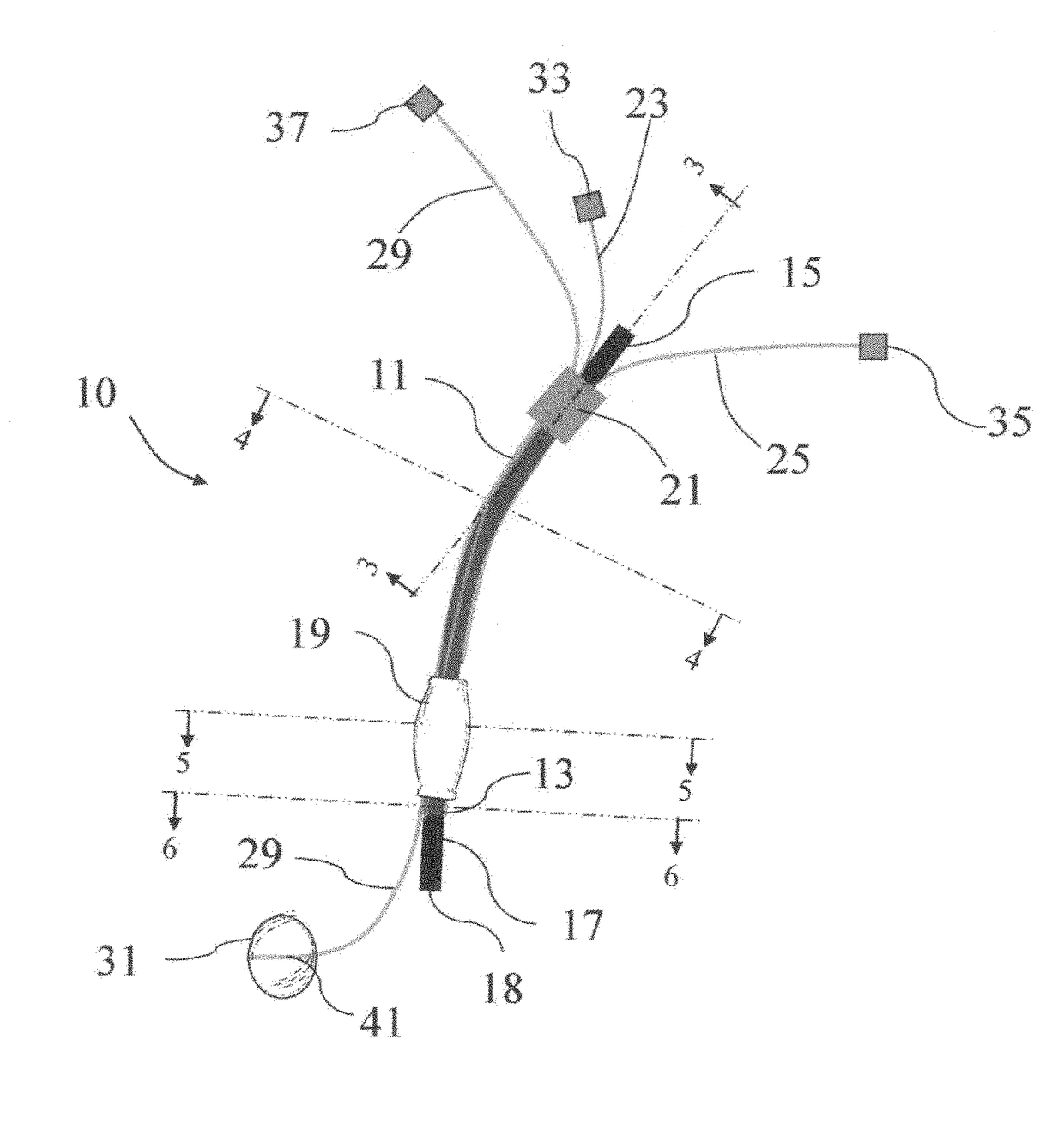
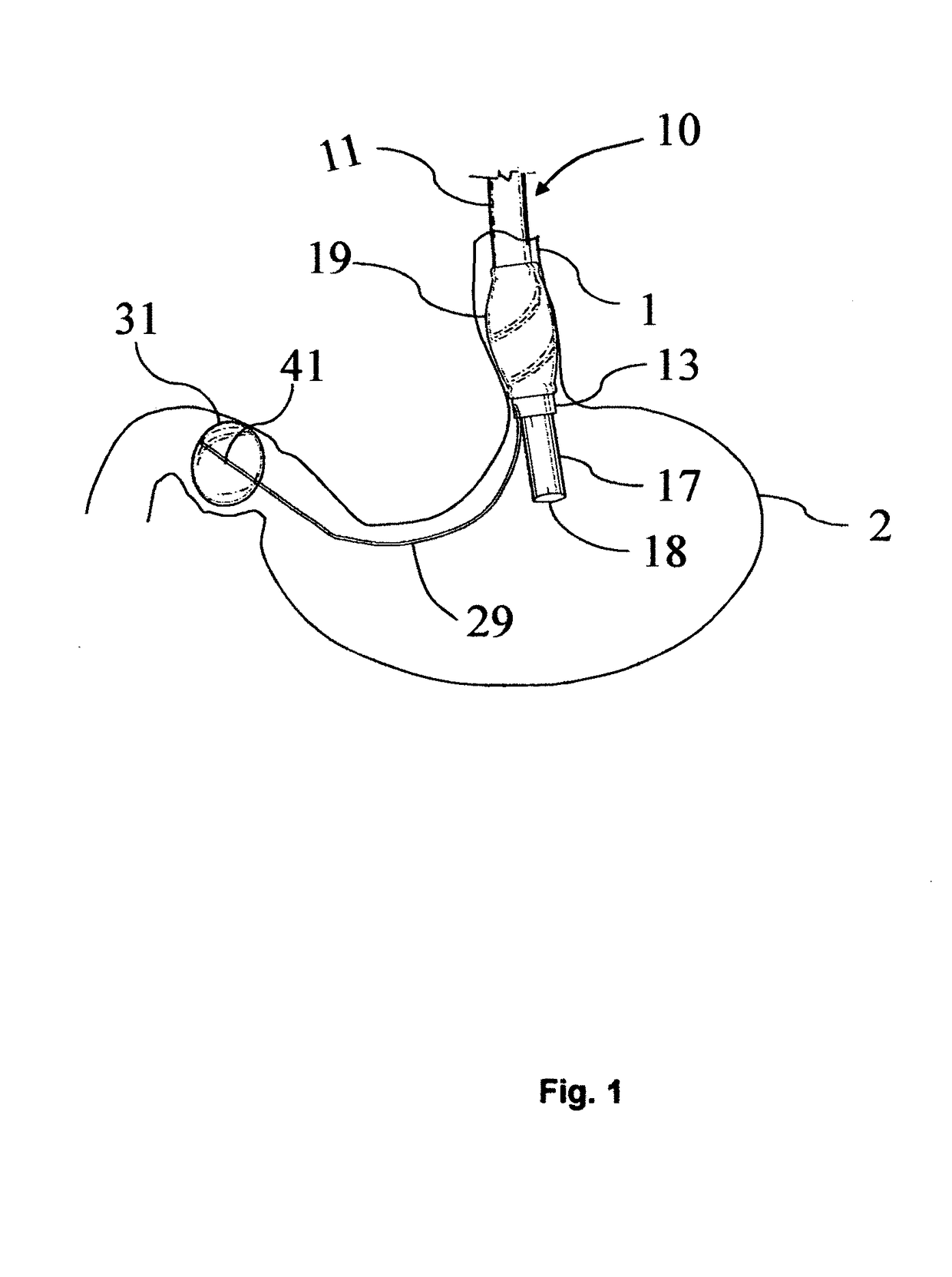
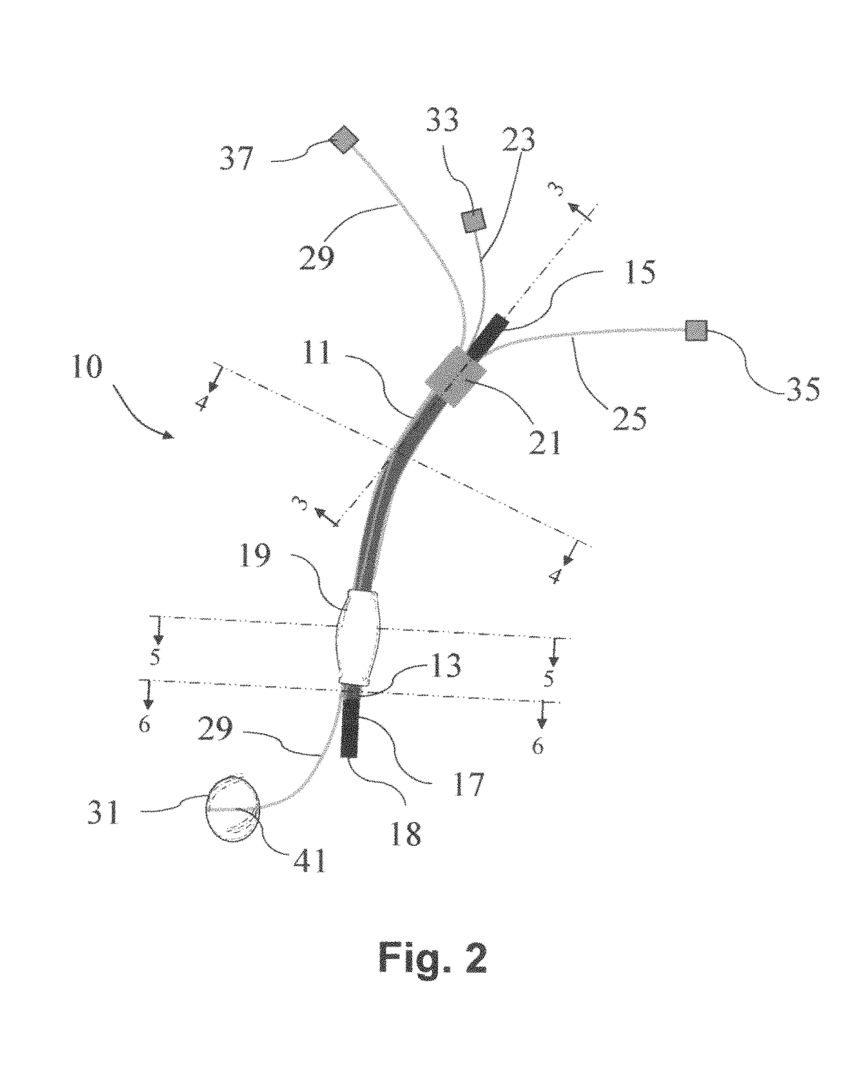
[0011]Double Balloon Endoscope Overtube 10 is made up of these components:

[0012]A—Overtube: As it is depicted in FIGS. 1 and 2, the Double Balloon Endoscope Overtube 10 is composed of a flexible tube 11 preferably transparent, that can be removably placed over a regular endoscope or echoendoscope shaft 17 and inserted inside a human gastrointestinal tract. The overtube has a proximal endportion 15 and a distal endportion 13. The overtube distal endportion 13 is inserted into the human gastrointestinal tract and an inflatable positioning balloon 19 is affixed to this area to secure the position of the overtube inside the body cavity. The overtube proximal endportion 15 stays out of the human body, and a cuff 21 is affixed to this portion to facilitate grasping and manipulation of the overtube 11 for insertion and positioning inside the body cavity by the endoscopist. The diameter of the overtube 11 is large enough to freeely receive a regular endoscope or echoendoscope shaft 17 there...


PUM


Abstract

This invention relates generally to an endoscope accessory. This device uses an overtube with an inflatable positioning balloon that can be inflated after placement of the overtube just proximal to the tip of the endoscope or echoendoscope. This balloon creates a temporary blockade of the gastrointestinal tract proximal to the portion that needs to be examined. The other part of the device is a catheter with an occlusion balloon affixed to its free endportion and can be passed through a passageway within the overtube and extended beyond the tip of the endoscope, distal to the part of the gastrointestinal tract that needs to be examined. Inflation of this balloon together with positioning balloon creates a closed space within the body cavity that can be filled with air or water for improving the quality of the examination with regular endoscope of echoendoscope, respectively, while reducing the examination complications.

Description

FIELD OF INVENTION[0001]This invention relates generally to an accessory for endoscopic examination of body organs particularly gastrointestinal tract. More particularly, the invention relates to creation of a closed region around the tip of an endoscope introduced into a visceral organ.BACKGROUND OF INVENTION[0002]An endoscope is a well-known optical system for evaluation of internal organs that was disclosed and claimed in U.S. Pat. No. 3,449,037 to C. J. Koester. Currently used fiberoptic endoscopes are comprised of many lenses mounted in a flexible tube to relay an image from inside a body cavity for viewing by a physicians for diagnosis or manipulation inside those cavitary spaces. Endoscopic ultrasound (EUS) is a device that combines endoscope and ultrasound to image the gastrointetinal wall and surrounding structures. The first prototype for human use was developed in 1980, and several generations of echoendoscopes have been developed since then. In the 1990 the capability of...

Claims


Application Information

Patent Timeline
Patent Type & Authority Patents(United States)
IPC IPC(8): A61B1/00A61B1/273A61B8/12A61B1/04A61M25/10A61B1/005
CPCA61B1/2736A61B1/00082A61B1/00135A61B8/12A61M2025/1052A61B1/005A61M25/1011
Inventor FARHADI, ASHKAN
Owner IZOMED INC
Why Eureka
Try Eureka