
Myeloid cell promoter and constructs containing same
Patent Information
- Authority / Receiving Office
- US · United States
- Current Assignee / Owner
- Publication Date
- 2005-04-19
- Estimated Expiration
- Not applicable · inactive patent
Smart Images
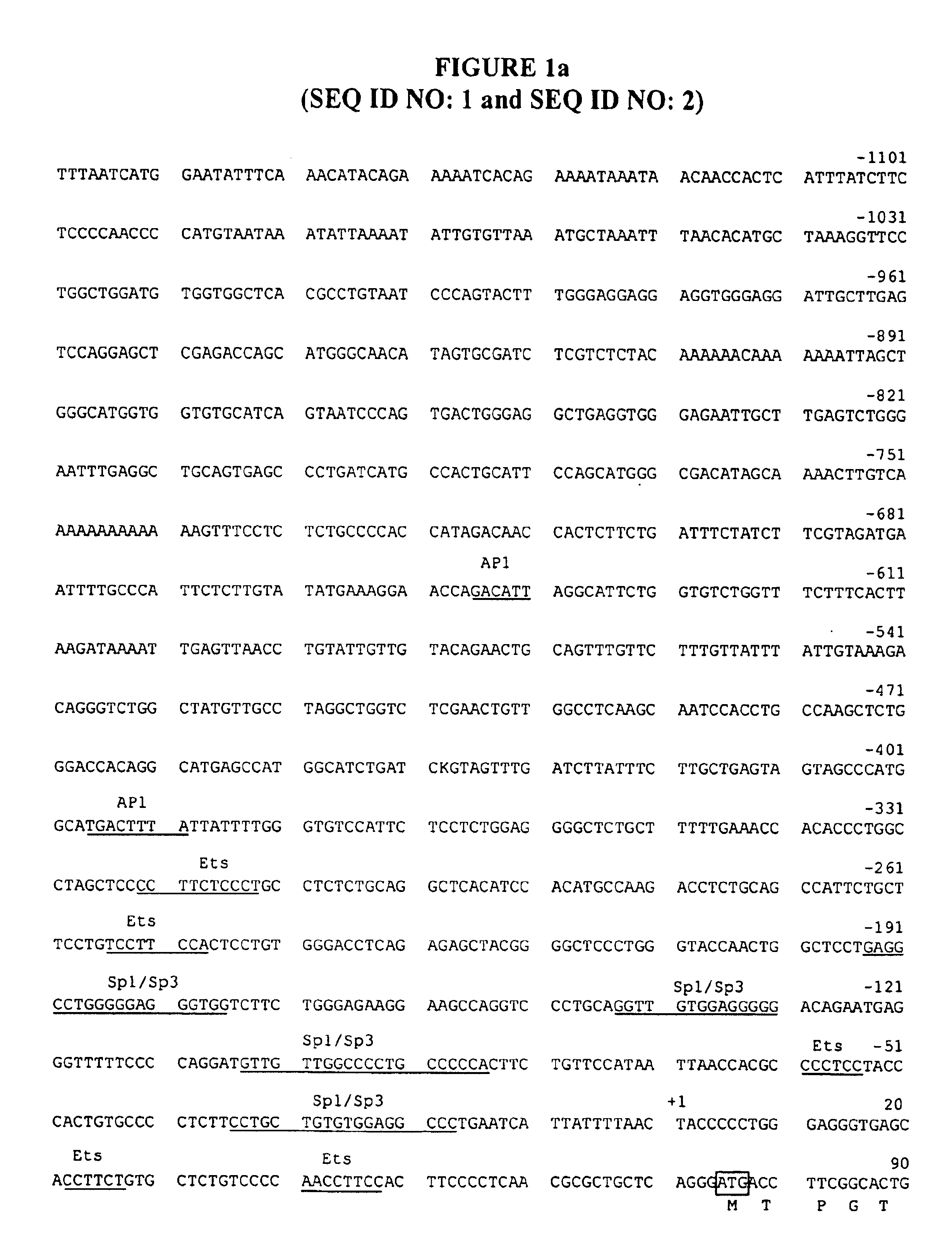
Figure 1 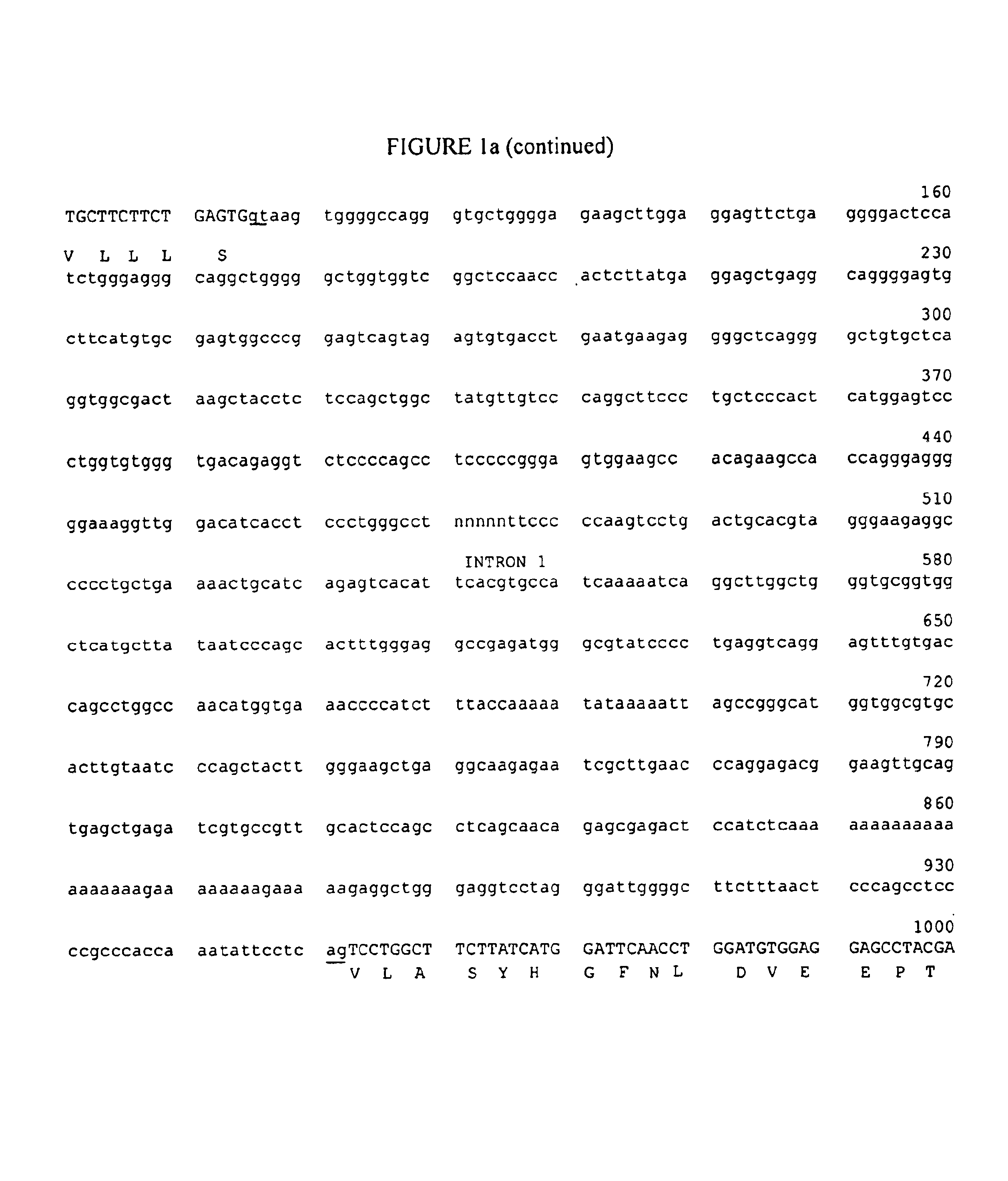
Figure 2 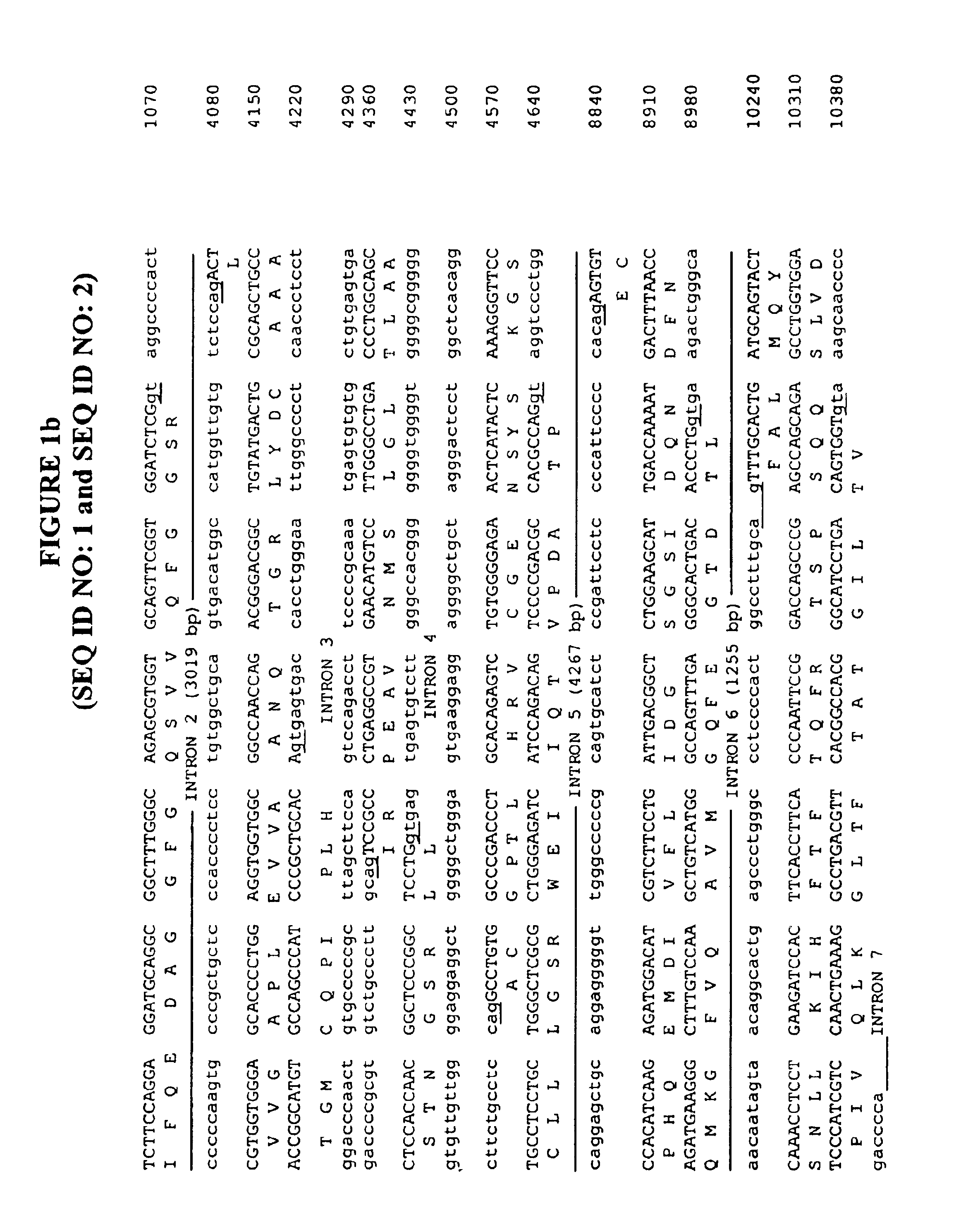
Figure 3
Abstract
Description
TECHNICAL FIELD
[0001] The present invention relates to a novel promoter isolated from a myeloid cell, as well as cis-acting elements from the myeloid cell, and to novel constructs containing the same.BACKGROUND OF THE INVENTION
[0002] The expression of foreign genes in various cell types has become a commonplace occurrence in the field of biotechnology. The expression of the foreign, or heterologous, gene is performed by the transcription of the genetic information in the gene from DNA to RNA. The RNA is then translated into the protein for which the gene encodes.
[0003] In transcription, the RNA is synthesized using the DNA of the gene for a template. The RNA is synthesized by a reaction catalyzed by RNA polymerases. The RNA polymerase binds to a particular molecular site on the DNA to initiate the transcription process. This site to which the RNA polymerase binds is known as the promoter.
[0004] Therefore, in order to express heterologous genes in foreign cells, the genes must be under t...
Examples
example 1
Methodologies and Materials For Identification of CD11d Promoter
[0070]The following methodologies and materials were used in the following Examples:
A. Cell Culture
[0071]The cell lines used were: THP-1 (acute monocytic leukemia, ATCC TIB-202), HL60 (promyelocytic leukemia, ATCC CCL 240), IM-9 (B-cell multiple myeloma, ATCC CCL-159), Jurkat (T-cell acute leukemia, ATCC TIB 152), MCF-7 (breast adenocarcinoma, ATCC HTB-22) and Schneider's Drosophila 2 (D. Melanogaster embryo, ATCC CRL 1963). THP-1, HL60, and Jurkat cells were grown in RPMI-1640 medium containing 10% fetal calf serum (Biofluids, Rockville, Md.). IM-9 cells were grown in RPMI-1640 medium containing 20% fetal calf serum (Biofluids, Rockville, Md.). HeLa and MCF-7 cells were grown in Dulbecco's modified Eagle's medium containing 10% fetal calf serum (Biofluids, Rockville, Md.). Drosophila Schneider 2 cells were grown in Schneider's medium containing 10% insect-tested fetal calf serum (Sigma, St. Louis, Mo.). All media conta...
example 2
Isolation and Sequence Analysis of The CD11d 5′ Flanking Region
[0084]The inventors isolated a genomic clone for CD11d that contains the intergenic region between CD11c and CD11d and the 5′-coding portion of CD11d. Utilizing this genomic clone, the regulatory mechanisms for CD11d expression were determined and the clone analyzed in order to locate the CD11d promoter and cis-acting elements. CD11d is positioned no more than 11.5 kb downstream of CD11c.
[0085]A collection of CD11c positive cosmid clones was re-screened for the presence of CD11d. One clone was completely sequenced and contains exons 15-30 of CD11c, and exons 1-6 of CD11d. Exon 7 was located separately on another clone. The genes were arranged in the same orientation, with the translational stop codon of CD11c positioned 11,461 bp upstream of the translational start codon of CD11d, as shown in FIG. 2. The sequence of the 1.2 kb 5′-flanking region and exons 1-7 are shown in FIG. 1.
[0086]DNA sequence analysis of this clone ...
example 3
Determination of the Transcriptional Start Site of the CD11d Gene
[0088]Poly (A+) RNA and total RNA from myeloid HL60 cells was subjected to primer extension analysis with primers N379 (antisense primer with 5′-end positioned 20 nucleotides downstream of the ATG start site) and N470 (antisense primer with 5′-end positioned immediately upstream of the ATG start site), respectively. The longest extension product obtained with N379 was 101 nucleotides long, positioning the transcriptional start site 78 nucleotides upstream of the ATG site. Two extension products, 92, and 93 nucleotides long, were observed with N470 total RNA and would position the transcriptional start site(s) 14 and 15 bp further upstream. No extension products were detected when total yeast RNA was analyzed.
[0089]An S1 nuclease protection assay was performed with a 247 nucleotide single-stranded DNA probe that was generated by extension of [γ-32P]ATP-labeled N470 primer on a double-stranded DNA template with exonuclea...