Automatic tissue calibration method for IVUS gray-scale image

A gray-scale image and automatic organization technology, applied in the field of medical imaging, can solve the problems of restricting wide application and achieve high repeatability

Active Publication Date: 2014-11-12
NORTH CHINA ELECTRIC POWER UNIV (BAODING)
View PDF3 Cites 13 Cited by
  • Summary
  • Abstract
  • Description
  • Claims
  • Application Information

AI Technical Summary

Problems solved by technology

However, not all IVUS imaging systems in clinical use allow the acquisition of raw radio frequency signals, and most existing equipment in hospitals requires hardware modification to use this technology, thus limiting the wide application of this method

Method used


Image

  • Automatic tissue calibration method for IVUS gray-scale image
  • Automatic tissue calibration method for IVUS gray-scale image
  • Automatic tissue calibration method for IVUS gray-scale image

Examples


Embodiment Construction

[0047] The method of the present invention obtains feature data of blood vessel wall tissue (including plaque tissue) by extracting texture features of IVUS gray-scale images, and uses an Adaboost classifier to complete automatic calibration of plaque tissue of different components after dimensionality reduction of the feature data. The steps of the inventive method are described in detail below:

[0048] 1. Extract the texture features of the IVUS grayscale image:

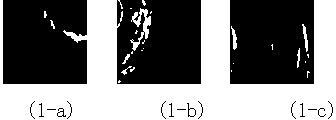
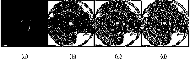
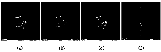
[0049] IVUS grayscale images do not contain color information, and due to the extremely fast image acquisition speed, the front and rear frames are very similar, so color features and shape features cannot be used as quantitative features for tissue calibration. However, IVUS grayscale images contain a large amount of texture information, and the texture difference between normal tissue and lesion tissue is obvious, so texture information can be used as an important basis for tissue calibration. IVUS gray-scale i...


PUM


Abstract

The invention discloses an automatic tissue calibration method for an IVUS gray-scale image. According to the technical scheme, the automatic tissue calibration method includes the steps of firstly, extracting textural features of the IVUS gray-scale image; secondly, carrying out dimension reduction processing on the extracted textural features; finally, calibrating textural feature data through an Adaboost classifier, and accordingly completing automatic identification and automatic calibration of different-ingredient plaque tissues. By means of the automatic tissue calibration method, original radio frequency signals and backscattering signals of an imaging device do not need to be collected, the calibration process is not limited by an IVUS imaging system and can be completed in a full-automatic mode, identification and calibration results which are objective, accurate and high in repeatability can be obtained, and the bases can be provided for computer-aided diagnosis of the coronary heart disease, making of an interventional treatment plan and the like.

Description

technical field [0001] The invention relates to a method for automatically performing vascular tissue calibration on intravascular ultrasonic grayscale images to distinguish normal blood vessel wall tissue from atherosclerotic plaque tissue with different components, and belongs to the technical field of medical imaging. Background technique [0002] Cardiovascular disease due to atherosclerosis is an inflammatory process affecting the walls of blood vessels, which will develop into a variety of plaques within the vessel lumen. Plaque tissue can be divided into four types according to its composition: lipid plaque, fibrotic plaque, calcified plaque and mixed plaque. Accurate detection and classification of plaque is of great significance for the diagnosis and treatment of atherosclerotic diseases. [0003] Intravascular ultrasound (IVUS) is an interventional imaging method commonly used in clinical diagnosis of vascular lesions and detection of vulnerable plaques. It inlay...

Claims


Application Information

Patent Timeline
Patent Type & Authority Applications(China)
IPC IPC(8): G06F19/00
Inventor 孙正王立欣周雅
Owner NORTH CHINA ELECTRIC POWER UNIV (BAODING)
Why Eureka
Try Eureka