

Probe set for diagnosing Xp11.2 ectopic perivascular epithelioid cell neoplasm and application of probe set
A technology of xp11.2 and perivascular, applied in the field of fluorescent in situ hybridization probe application, can solve the problems such as PSF-TFE3 fusion gene has not been reported, achieve high success rate and improve the effect of accuracy
- Summary
- Abstract
- Description
- Claims
- Application Information
AI Technical Summary
Problems solved by technology
Method used
Image


Examples
Embodiment 1
[0029] Embodiment 1: the preparation of DNA probe combination:
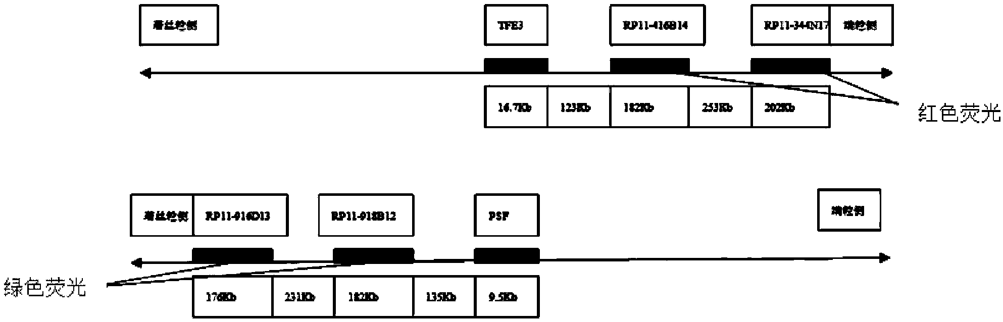
[0030] Select two BAC clone fragments that can be connected at the telomeric side of the TFE3 gene of chromosome X and the centromere side of the PSF gene of chromosome 1, and control the farthest distance between the probes at both ends within 1500kb, and keep a certain distance between the BAC clone fragments. The distances do not overlap, and the fragments are of similar size. The cloned fragments were obtained from the Human BAC Cloning Center of EmpireGenomics (http: / / www.empiregenomics.com / helixhq / clonecentral / search / human). The BAC clone fragments on the centromere side of PSF are RP11-918B12 (fragment length 182kb) and RP11-916D13 (fragment length 176kb), and the BAC clone fragments on the telomeric side of TFE3 are RP11-416B14 (fragment length 182kb) and RP11-344N17 ( Fragment length 202kb). The linking sequence of BAC cloned fragment and PSF gene is PSF, RP11-918B12, RP11-916D13, centromere of chromos...
Embodiment 2
[0032] Embodiment 2: fluorescence in situ hybridization process:
[0033] 1. Tissue microarray construction of specimens:
[0034] 61 cases of PEComa diagnosed in Nanjing General Hospital of Nanjing Military Region were collected. Two experienced pathologists referred to the WHO soft tissue tumor classification criteria, immunohistochemical TFE3 positive, and RT-PCR to detect fusion gene results ( figure 2 , one of the 61 patients was diagnosed at the mRNA level, and the correspondence with the results of this experiment can better reflect the reliability of this experiment), and the diagnostic evaluation was performed, and the final diagnosis of Xp11.2 translocation PEComa was in 5 cases, and other Type PEComa in 56 cases. The above specimens (including tumor and adjacent tissue) were made into tissue chips ( image 3 ), all specimens were fixed in 10% formalin, processed routinely, and embedded in paraffin. Tissue microarrays were fabricated by hand. The tissue microar...
Embodiment 3
[0047] Example 3: Xp11.2 Translocation PEComa Diagnostic Kit
[0048] The kit contains the probe combination described in Example 1, and the characteristics of the probe combination are mainly:
[0049] (1) The BAC clone probes on the centromere side of PSF are RP11-918B12 (fragment length 182kb) and RP11-916D13 (fragment length 176kb), labeled with green fluorescence. These two probes are a green fluorescent signal under the fluorescent microscope, representing the centromere side of PSF gene.
[0050] (2) The BAC clone probes on the telomeric side of TFE3 are RP11-416B14 (fragment length 182kb) and RP11-344N17 (fragment length 202kb), labeled with red fluorescence. These two probes show a red fluorescent signal under the fluorescent microscope, representing the telomeric side of the TFE3 gene.
[0051] (3) Under normal circumstances, the red and green signals are separated. In Xp11.2 translocation PEComa, due to the fusion of the X chromosome TFE3 gene and the No. 1 chromo...
PUM
 Abstract
Description
Claims
Application Information