

Simulator model used for simulating tumor ultrasonic radiography
A technology of contrast-enhanced ultrasound and ultrasound contrast agent, which is applied in the field of phantom models, can solve the problems of distinguishing and unusable normal tissue models, and achieve the effects of easy acquisition, controllable consistency of finished products, and simple materials
- Summary
- Abstract
- Description
- Claims
- Application Information
AI Technical Summary
Problems solved by technology
Method used
Image

Examples
Embodiment 1
[0076] The preparation of embodiment 1 phantom model
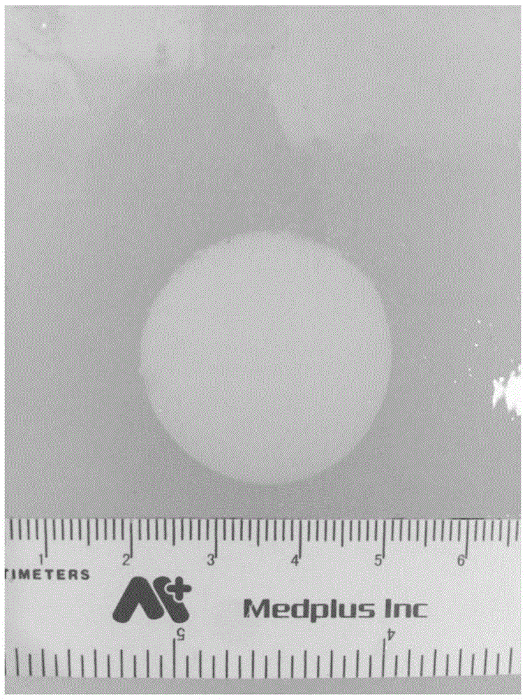
[0077] Weigh 20g of carrageenan and 1g of gastric window acoustic contrast agent into a beaker with an electronic balance, add 1000mL of double distilled water, stir with a glass rod until uniform, heat, and place the beaker in a constant temperature water bath at 60°C to maintain the temperature. A normal tissue glue solution is formed. Take 38mL of normal tissue glue in a small beaker, add 2mL of milk to form tumor glue Matrigel, then add lipid microbubble ultrasound contrast agent SonoVue (Bracco, Milan, Italy), stir evenly to form tumor glue. In this example, 6 ml of physiological saline was added to each bottle of SonoVue to dissolve as a mother solution, and tumor gel models with SonoVue concentrations of 0.07%, 0.105%, 0.1225%, 0.14%, 0.175% and 0.21% (w / v) were prepared respectively. After placing the tumor glue in a sphere mold with a diameter of 3 cm, place it in ice water and cool it for 5 minutes, and take it ...
Embodiment 2
[0079] Embodiment 2 Ultrasonic imaging effect detection of phantom model
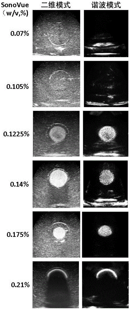
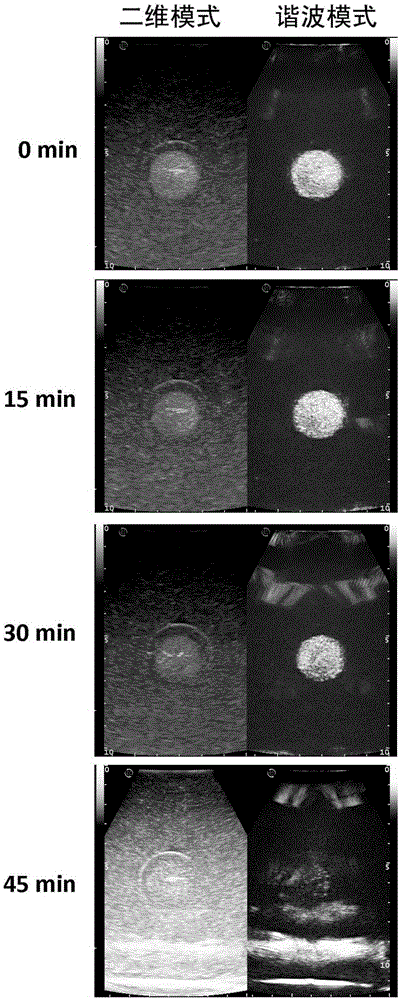
[0080] Phantom phantoms with different SonoVue concentrations were examined with CEUS. In this embodiment, Esaote MyLabtwice model clinical ultrasonic diagnostic instrument and linear array probe LA332 are used to detect the phantom, the imaging frequency is 3-11 MHz, the focal point is placed in the middle of the tumor, and the gain is 0.5. For harmonic contrast ultrasound imaging mode, use a mechanical index of 0.03 and a gain of 0.38. 10 min after the phantom model was taken out of the refrigerator was defined as 0 min, and two-dimensional ultrasound and harmonic contrast ultrasound imaging were performed on the phantom model at different time points.
[0081] The result is as figure 2 As shown, at the time point of 0 min, phantom models prepared with different concentrations of SonoVue presented different imaging effects. When the concentration of SonoVue was low, the tumor glue and normal tissue...
Embodiment 4
[0086] Embodiment 4 phantom model preparation temperature control
[0087] When making the phantom model, the temperature of the solution is adjusted within a certain range of 50-70°C. As shown in Table 1. When the temperature is 50°C, the echogenicity of the tumor glue fades the latest, but the solution is easy to solidify at this time, so the preferred temperature is about 60°C, for example, 57-65°C. Trainers can adjust the temperature of the water bath according to their own needs and make corresponding adjustments.
[0088] Table 1. Imaging time of tumor gels with different SonoVue contents at different temperatures
[0089]
PUM
 Abstract
Description
Claims
Application Information