Immunochromatographic assay test paper strip for quantitatively detecting carcino-embryonic antigen (CEA) and preparation method thereof

A technology of immunochromatographic test paper and carcinoembryonic antigen, which is applied in the field of medical detection, and achieves the effects of simple operation, convenient and fast inspection, and improved sensitivity

Inactive Publication Date: 2016-08-24
SHANGHAI INST OF MICROSYSTEM & INFORMATION TECH CHINESE ACAD OF SCI
View PDF3 Cites 2 Cited by
  • Summary
  • Abstract
  • Description
  • Claims
  • Application Information

AI Technical Summary

Problems solved by technology

LFIA technology is based on immunolabeled nanoparticles, such as gold nanoparticles, quantum dots, etc., but the sensitivity of immuno

Method used


Image

  • Immunochromatographic assay test paper strip for quantitatively detecting carcino-embryonic antigen (CEA) and preparation method thereof
  • Immunochromatographic assay test paper strip for quantitatively detecting carcino-embryonic antigen (CEA) and preparation method thereof
  • Immunochromatographic assay test paper strip for quantitatively detecting carcino-embryonic antigen (CEA) and preparation method thereof

Examples


Embodiment 1

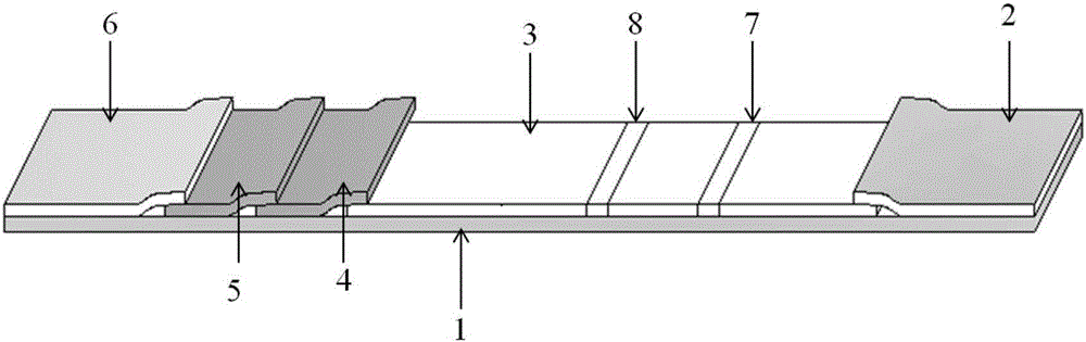
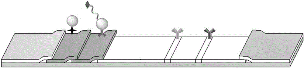
[0030] Such as figure 1 As shown, this embodiment provides an immunochromatographic test strip for the quantitative detection of carcinoembryonic antigen, including a bottom plate 1, an absorbent pad 2, a nitrocellulose membrane 3, a first binding pad 4, a second binding pad 5 and a sample Pad 6. The first binding pad 4 is coated with detection magnetic beads, and the second binding pad 5 is coated with signal magnetic beads. IgG antibody (C line) 7 and CEA coated antibody (T line) 8 are sprayed on the nitrocellulose membrane 3 .

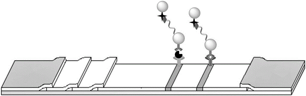
[0031] The serum sample is added dropwise to the sample pad, and the liquid flows through the second binding pad, the first binding pad, the T line and the C line in sequence. When the sample contains CEA antigen, the antigen first binds to the detection magnetic beads on the first binding pad, and when the liquid flows forward to the T line due to chromatography, the coated antibody can also recognize the CEA antigen, forming a "double antibody ...

Embodiment 2

[0033] 1) Coating nitrocellulose membrane: use sterile sodium chloride solution with a concentration of 0.135M to dilute the IgG antibody to a concentration of 1mg / ml; use PBS buffer with a concentration of 10mM and pH=7.4 to coat CEA with the antibody The concentration was diluted to 2mg / ml. First, assemble the nitrocellulose membrane at a suitable position on the bottom plate, and then use a quantitative liquid spray device to spray two kinds of antibodies on different positions of the nitrocellulose membrane to form C lines and T lines. The plain film was dried at 25°C for 4 hours, and stored in a dry environment.

[0034]2) Pretreatment of the sample pad: soak the glass cellulose membrane in the sample pad treatment solution, take it out after about 30 minutes, dry it at 37°C for 4 hours, and store it in a dry environment. The formula of described sample pad treatment liquid is: containing 0.003M boric acid, 0.004M sodium borate, 0.5% (w / v) sodium chloride, 0.5% (w / v) bov...


PUM


Abstract

The invention relates to an immunochromatographic assay test paper strip for quantitatively detecting a carcino-embryonic antigen (CEA) and a preparation method thereof. The immunochromatographic assay test paper strip mainly comprises a sample pad, a first combining pad, a second combining pad, a cellulose nitrate membrane, a bottom plate and an absorbent paper, wherein the sample pad is treated, the first combining pad is coated with detection magnetic beads, and the second combining pad is coated with signal magnetic beads; an IgG antibody, i.e., a quality control line and a CEA coated antibody, i.e., a detection line are fixed on the cellulose nitrate membrane. The preparation method comprises the step of sequentially stacking and combining the absorbent paper, the cellulose nitrate membrane, the combining pads and the sample pad. According to the immunochromatographic assay test paper strip for quantitatively detecting the CEA and the preparation method thereof, characteristics of the magnetic beads and characteristics of an amplifying system are integrated, the aims of specific sensitivity and quantitative detection are achieved, and the cost is low; the immunochromatographic assay test paper strip simultaneously supports detection on small samples and large samples, is simple in operation and convenient and rapid in detection and is suitable for being subjected to large-scale popularized production.

Description

technical field [0001] The invention belongs to the field of medical detection, in particular to an immunochromatographic test strip for quantitative detection of carcinoembryonic antigen and a preparation method thereof. Background technique [0002] Carcino-embryonic antigen (CEA) is a glycoprotein on the surface of cells, and a high CEA content can usually be detected in developing embryonic tissues. The content of CEA in normal adult serum is very low, but the level of CEA in various cancer patients is abnormally elevated. CEA is closely related to the spread and metastasis of tumors, and it has been clinically used as a broad-spectrum glycoprotein tumor marker, widely used in the detection of gastrointestinal cancer, lung cancer, intestinal cancer and pancreatic cancer, etc. The distinction between tumors and malignant tumors and the judgment of tumor staging. Some non-tumor diseases, such as inflammation and polyps, will increase the serum CEA content to a certain ex...

Claims


Application Information

Patent Timeline
IPC IPC(8): G01N33/574G01N33/558
CPCG01N33/57473G01N33/558
Inventor 毛红菊刘芳铭孔祥燕张宏莲金庆辉赵建龙
Owner SHANGHAI INST OF MICROSYSTEM & INFORMATION TECH CHINESE ACAD OF SCI
Why Eureka
Try Eureka