Cerebral stroke CT image segmentation method

A technique for CT imaging, stroke

Active Publication Date: 2021-07-27
CENT SOUTH UNIV
View PDF4 Cites 6 Cited by
  • Summary
  • Abstract
  • Description
  • Claims
  • Application Information

AI Technical Summary

Problems solved by technology

At the same time, most of the attention mechanisms used in the existing CT-based infarct segmentation methods are channel attention mechanisms, which cannot handle the spatial location attention mechanism to simulate the spatial location continuity of infarction, and the accuracy is low.

Method used


Image

  • Cerebral stroke CT image segmentation method
  • Cerebral stroke CT image segmentation method
  • Cerebral stroke CT image segmentation method

Examples


Embodiment Construction

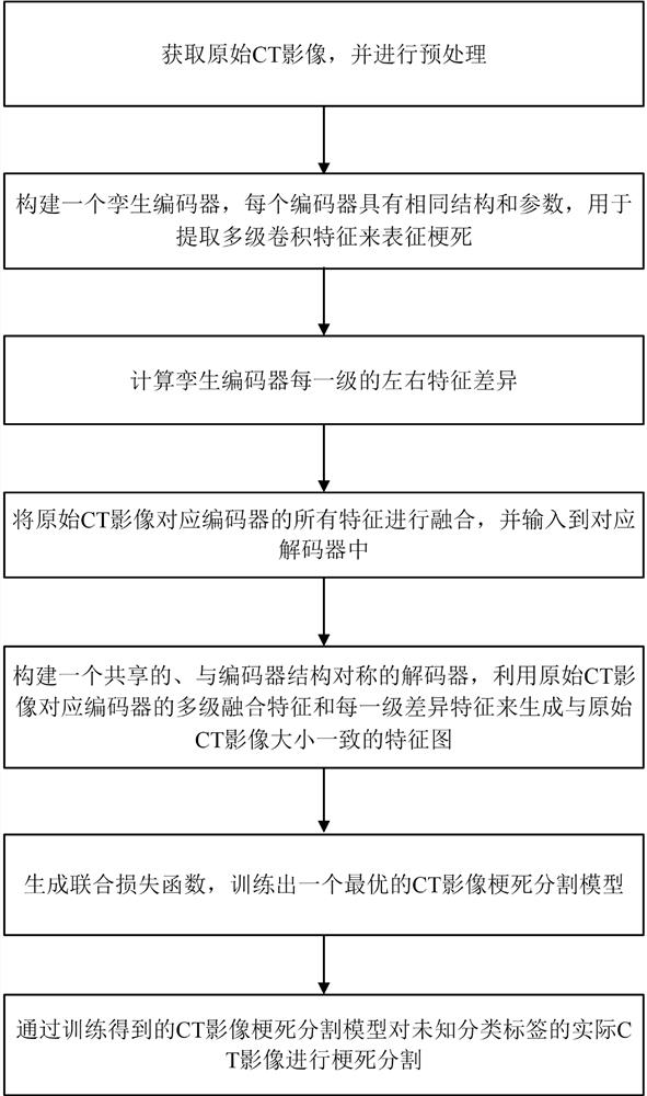
[0069] like figure 1 It is a schematic flow chart of the method of the present invention: the stroke CT image segmentation method provided by the present invention includes the following steps:

[0070] S1. Obtain the original CT image, and perform cross-sectional left-right flip and registration of each obtained brain CT image to obtain the flip CT image, and process the original CT image and the flip CT image;

[0071] S2. Construct a twin encoder, each encoder has the same structure and parameters, and extract multi-level convolutional features from the original CT image and flipped CT image to characterize the infarction;

[0072] S3. For each level of the twin encoder, use the feature difference calculation module to obtain the left and right feature difference of each level of the CT image;

[0073] S4. Use the multi-level fusion module to fuse all the features of the corresponding encoder of the original CT image and input it into the corresponding decoder;

[0074] ...


PUM


Abstract

The invention discloses a cerebral stroke CT image segmentation method, which comprises the following steps of: overturning an ischemic cerebral stroke CT image, and preprocessing an original CT image and the overturned CT image; constructing twin multistage encoders, calculating the feature difference of each stage of the two encoders by a calculation module, and fusing features by using a multistage feature fusion module; constructing a shared decoder; designing a joint loss function, and training an optimal segmentation model on the training set; and finally, performing ischemic cerebral apoplexy infarction segmentation on a test set of an unknown segmentation label by using the trained segmentation model. According to the invention, the feature difference calculation module is used to calculate the feature difference of each level of two encoders, and the multi-level feature fusion module is used to fuse the global and local features; the infarction in the CT image can be accurately segmented, and technical support and reference are provided for improving the ischemic stroke diagnosis efficiency and accuracy and reducing the fatality rate and disability rate.

Description

technical field [0001] The invention belongs to the technical field of image data processing, and in particular relates to a stroke CT image segmentation method. Background technique [0002] Stroke seriously endangers the health of Chinese people. Acute ischemic stroke accounts for about 70% of stroke in my country and is the most common type of stroke. Due to the short treatment time window of ischemic stroke, computed tomography (CT) with shorter acquisition time and lower cost has become the preferred imaging technique in clinical ischemic stroke. The volume of infarct lesions in ischemic stroke is an important indicator for assessing the severity of stroke and decision-making for treatment. Currently, experts generally perform infarct segmentation on CT images manually. However, infarct segmentation by experts is very time-consuming and there are large subjectivity and individual differences. Therefore, more and more researchers pay attention to the automatic ischemi...

Claims


Application Information

Patent Timeline
Patent Type & Authority Applications(China)
IPC IPC(8): G06T7/00G06T7/11G06T7/33G06K9/46G06K9/62G06N3/04G06N3/08
CPCG06T7/0012G06T7/11G06T7/33G06N3/08G06T2207/10081G06T2207/20076G06T2207/20081G06T2207/20084G06T2207/30016G06V10/44G06N3/047G06N3/048G06N3/045G06F18/2415G06F18/241
Inventor 匡湖林刘锦王建新
Owner CENT SOUTH UNIV
Why Eureka
Try Eureka