
Method and System for Wavelet Based Detection of Colon Polyps
a wavelet and polyp technology, applied in the field of wavelet based detection of colon polyps, can solve the problems of large amount of data in digital images that it is difficult and tedious for a human, such as a physician, to interpret without additional aid, and existing methods generally do not detect the characteristic symptoms of various cancers
- Summary
- Abstract
- Description
- Claims
- Application Information
AI Technical Summary
Benefits of technology
Problems solved by technology
Method used
Image

Examples
first embodiment
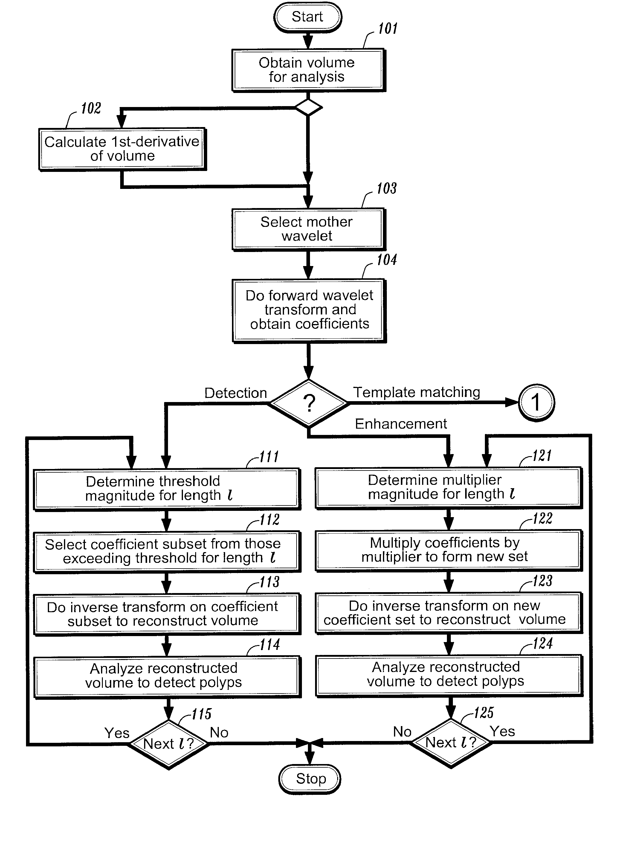
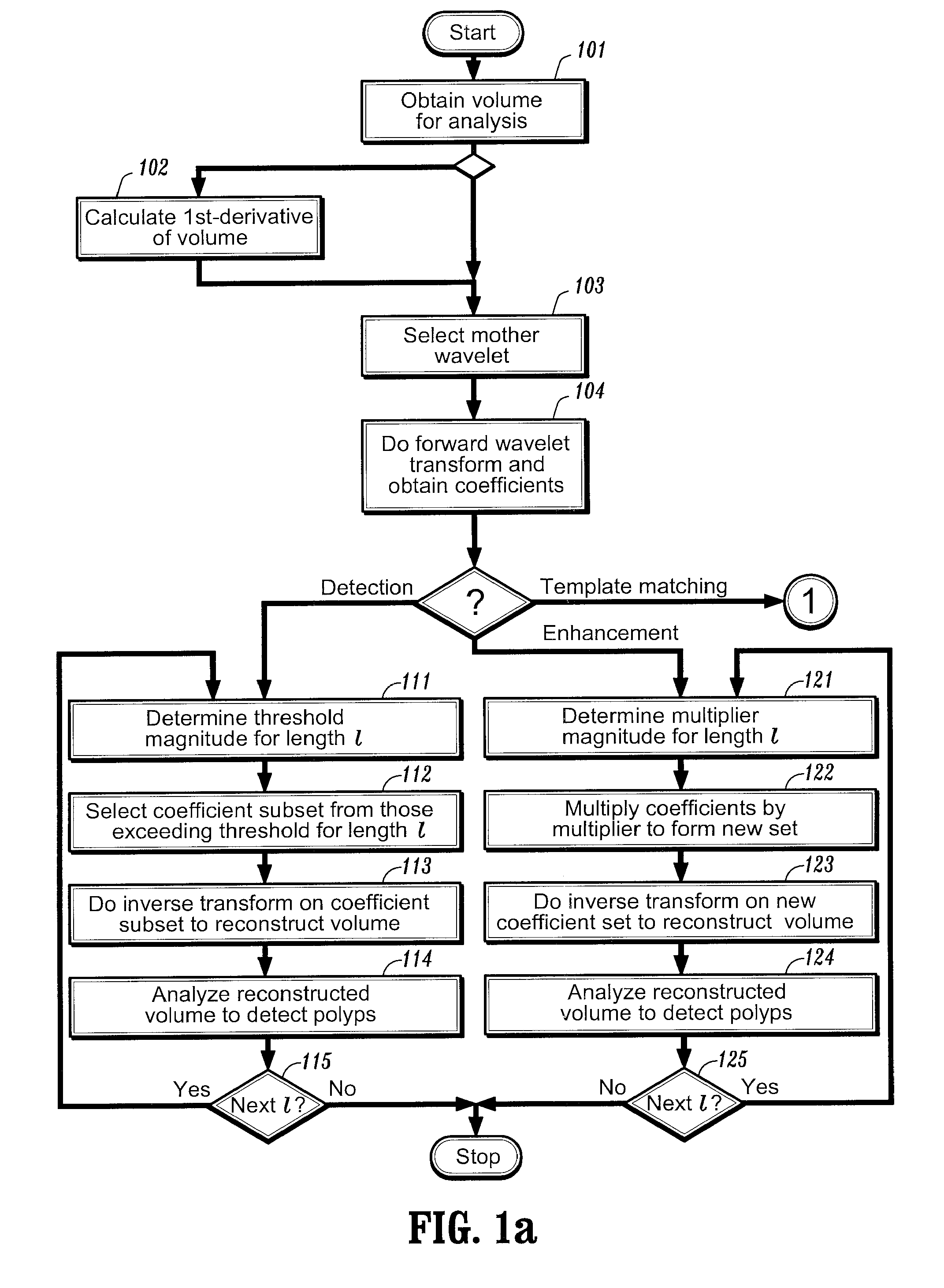
[0027] After preprocessing the image, one can perform a forward wavelet decomposition of a volume of data points via, e.g., the lifting scheme, using an appropriate mother scaling function. Although the volume being analyzed can be an image volume, it need not be so and the volume data points can have other meanings, as will be discussed below. For ease of explanation, the embodiments described herein below will be described in terms of an image. Referring now to FIG. 1a, after obtaining a volume of data points for analysis at step 101, one then selects a mother wavelet function at step 103, and performs the forward wavelet transform on the data volume at step 104. A first embodiment of the invention can be used for the detections of polyps and other structures of interest. Assuming one started with an image comprising n points, one ends up with a set of n coefficients of varying length scales. A wavelet basis function whose shape and length scale correspond to a polyp (or cavity) w...
second embodiment
[0030] In a second embodiment of the invention, after suitable preprocessing, one can again start by performing a forward wavelet decomposition of an image of n points to obtain n basis functions and associated coefficients.
[0031] In this embodiment, rather than thresholding the coefficients, one determines at step 121 a fixed multiplier value that is dependent on scale: γ′1=2−1αγ1, where α is a constant multiplier coefficient. The wavelet coefficients are multiplied by this multiplier at step 122. As with the first embodiment, for the first few times that this procedure is applied for detecting a polyp, α will be determined by inspection. However, once an appropriate value of α has been found, that value can be automatically applied in future enhancement procedures. One can then reconstruct the volume at step 123 by using the inverse wavelet transform on the primed set of coefficients. The resulting volume would contain those regions suspicious of being a polyp appearing enhanced ...
third embodiment
plate Matching
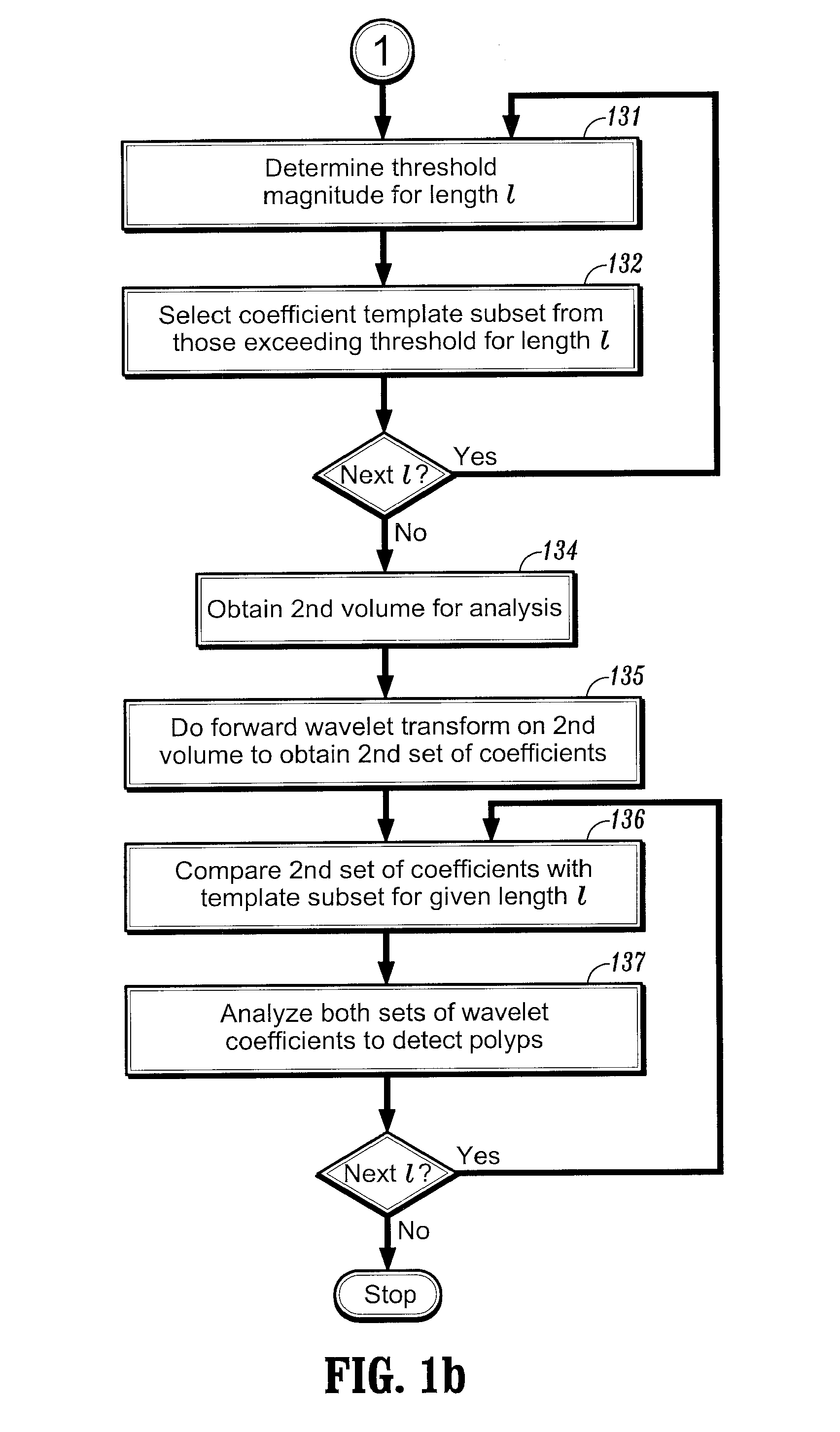
[0032] In the third embodiment of invention, one seeks to detect a polyp in the wavelet domain rather than the spatial domain. Once again, after suitable preprocessing, one can start by performing a forward wavelet decomposition of an image of n points to obtain n basis functions and associated coefficients. Turning now to FIG. 1b, as in the first embodiment, coefficient thresholds can be defined at step 131 for each scale coefficient l based on the intensity: γth=2−1β, where β represents a threshold intensity value, and that subset of m coefficients exceeding the threshold γth is then selected at step 132. This process is repeated to select a subset of coefficients Tl for each length scale l, and can also be repeated for differently shaped basis functions.
[0033] These subsets of coefficients can then be used as templates for detecting polyps as follows. Given another input image volume at step 134, one can apply the forward wavelet transform to obtain another set of ...
PUM
 Abstract
Description
Claims
Application Information