Method of processing radiological images, and, in particular, mammographic images

a radiological image and mammogram technology, applied in the field of radiological image processing, can solve the problems of inconvenient recombination technique, variation in the grey level appearing in the recombined image, and breasts with a uniform thickness, etc., and achieve the effect of improving quality and reducing the non-uniform background area of the recombined imag

Active Publication Date: 2009-12-10
GENERAL ELECTRIC CO
View PDF8 Cites 18 Cited by
  • Summary
  • Abstract
  • Description
  • Claims
  • Application Information

AI Technical Summary

Benefits of technology

[0027]An embodiment of the invention may be configured to produce a recombined image of improved quality, i.e., wherein the nonuniformities of the background area of the recombined image have been reduced, or even eliminated.

Problems solved by technology

However, in actual practice, this recombination technique is not suitable when the radiation energy spectra used are not monoenergetic.
In addition, other factors are also the cause of variations in the grey levels appearing in the recombined image.
As a matter of fact, despite being compressed, the breast does not have a uniform thickness.
These variations in grey level are troublesome for the radiologist, because they make it difficult to view the outer edge portion of the breast without changing the windowing level used to view the interior of the breast (i.e., the compressed area of the breast)
Furthermore, these variations in grey level are likely to be interpreted as signs of the presence of a contrast medium and to result in image interpreting errors.

Method used


Image

  • Method of processing radiological images, and, in particular, mammographic images
  • Method of processing radiological images, and, in particular, mammographic images
  • Method of processing radiological images, and, in particular, mammographic images

Examples


Embodiment Construction

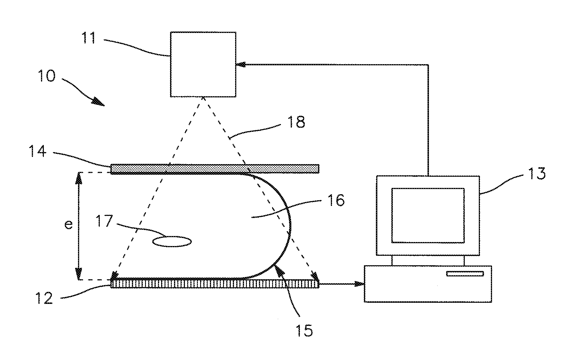
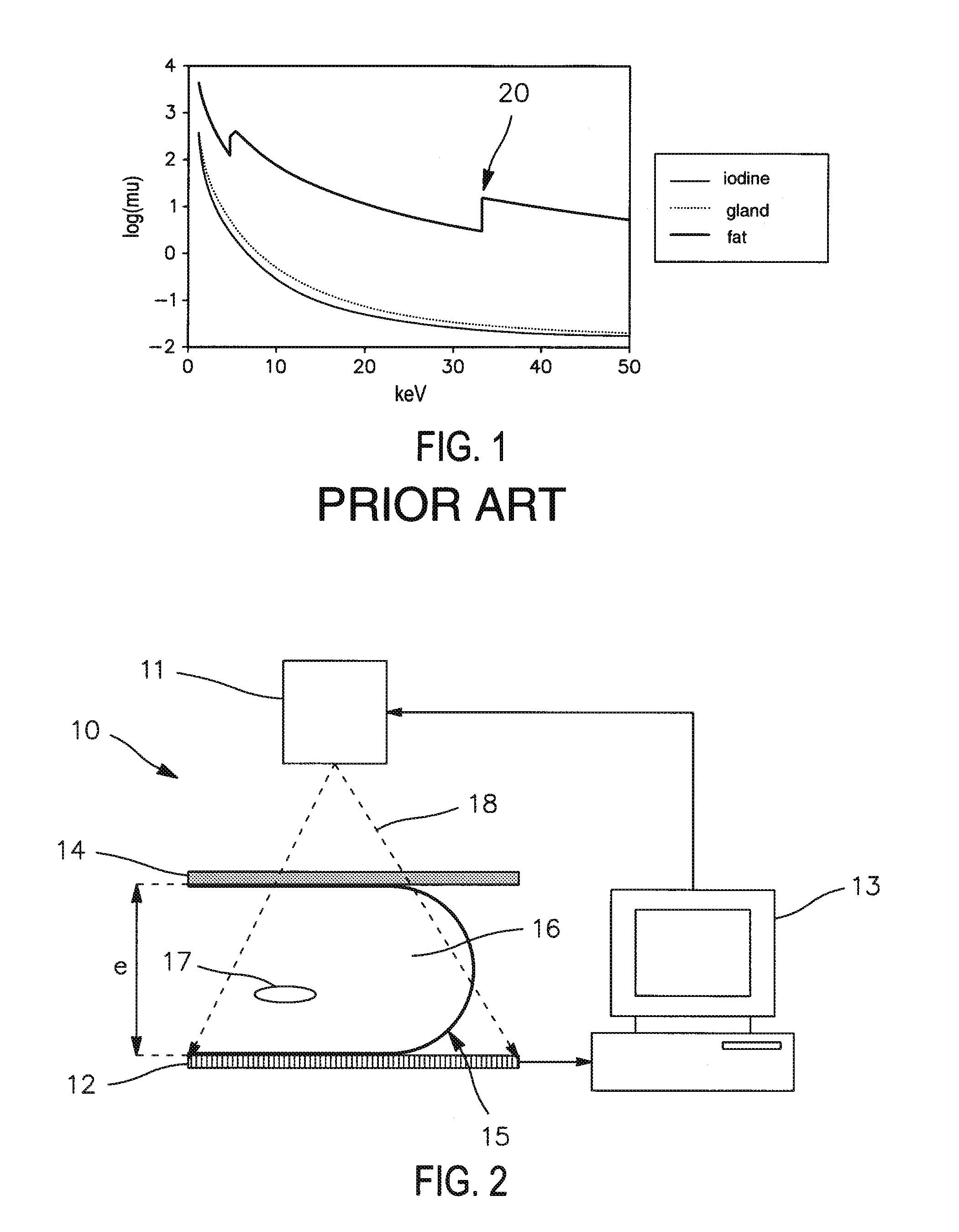
[0059]FIG. 2 is a schematic representation of a mammographic image acquisition and processing device 10.

[0060]The device 10 includes a source 11 capable of emitting X-rays 18, a digital detector 13 capable of receiving and detecting the rays emitted by the source 11, and a processing unit 13 capable of controlling the source 11 and of receiving and processing images acquired by the detector 12.

[0061]One breast 15 of a patient is arranged between the source 11 and the detector 12.

[0062]The device 10 also includes a compression plate 14 for compressing the breast 15 so that the breast has a compressed portion of substantially constant thickness.

[0063]Prior to this, the breast 15 received an injection of a contrast medium. The breast 15 includes an area of tissue 16 devoid of any contrast medium and one or more areas 17 of tissue in which the contrast medium has accumulated.

[0064]The processing unit 13 is capable of controlling the source 11 in order to vary the energy spectrum of the ...


PUM


Abstract

A method of processing a radiological image of an organ. The method may comprise generating a recombined image (R) from at least one previously acquired low energy image (L) and one high energy image (H) of a portion of the organ. The recombined image comprises a background and one or more areas corresponding to the presence of a contrast medium in the organ. The method may further comprise correcting the recombined image (R) by compensating (C) for the nonuniformities present in the background to obtain a compensated recombined image in which the background has a substantially uniform grey level.

Description

CROSS-REFERENCE TO RELATED APPLICATIONS[0001]This application claims priority under 35 U.S.C. §119(a)-(d) or (f) to prior-filed, co-pending French patent application serial number 0853763, filed on 6 Jun. 2008, which is hereby incorporated by reference in its entirety.STATEMENT REGARDING FEDERALLY SPONSORED RESEARCH OR DEVELOPMENT[0002]Not ApplicableNAMES OF PARTIES TO A JOINT RESEARCH AGREEMENT[0003]Not ApplicableREFERENCE TO A SEQUENCE LISTING, A TABLE, OR COMPUTER PROGRAM LISTING APPENDIX SUBMITTED ON COMPACT DISC[0004]Not ApplicableFIELD OF THE INVENTION [0005]The field of the invention relates to a method of processing radiological images, and, in particular, mammographic images.Description of Related Art[0006]In the field of radiology, and more particularly in mammography, image acquisition techniques referred to as “dual energy” are known. These techniques consist in injecting a contrast medium (e.g., containing iodine) into tissues in a region of the body being imaged (e.g.,...

Claims


Application Information

Patent Timeline
Patent Type & Authority Applications(United States)
IPC IPC(8): G06K9/00A61B6/04
CPCA61B6/583G06T5/50
Inventor PUONG, SYLVIEIORDACHE, RAZVANBOUCHEVREAU, XAVIERMULLER, SERGE LOUIS WILFRID
Owner GENERAL ELECTRIC CO
Why Eureka
Try Eureka