Ultrasound imaging apparatus and method for segmenting anatomical objects

Active Publication Date: 2017-12-21
KONINKLJIJKE PHILIPS NV
View PDF10 Cites 0 Cited by
  • Summary
  • Abstract
  • Description
  • Claims
  • Application Information

AI Technical Summary

Benefits of technology

[0008]It is an object of the present invention to provide an improved ultrasound imaging apparatus which can segment anatomical objects in ultrasound data with high precision and low technical effort. It is further an object of the present invention to p

Problems solved by technology

However a majority of the known segmentation techniques have a reduced performance for ultrasound imaging systems, since the segmentation model cannot properly be aligned to the ultrasound images which have a poor signal-to-noise ratio.
Usually a prior information about the shape of the anatomical structure to be segmented can improve the segmentation of the anatomical structures in the ultrasound images and these techniques are typically applied to three-dimensional data which require a large computation effort so that these techniques are typically expensive and cannot be applied to real-time applicat

Method used


Image

  • Ultrasound imaging apparatus and method for segmenting anatomical objects
  • Ultrasound imaging apparatus and method for segmenting anatomical objects
  • Ultrasound imaging apparatus and method for segmenting anatomical objects

Examples


Embodiment Construction

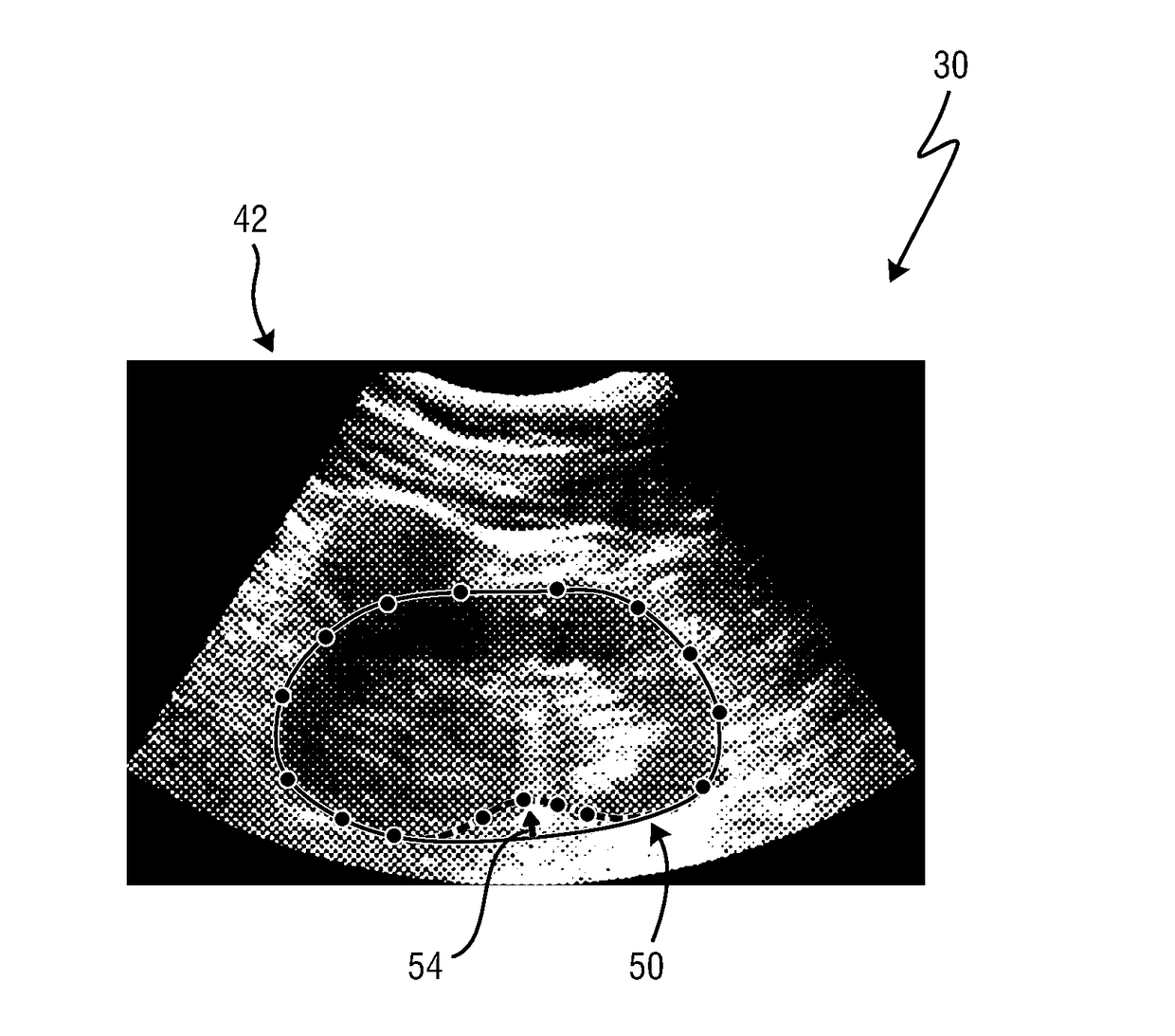
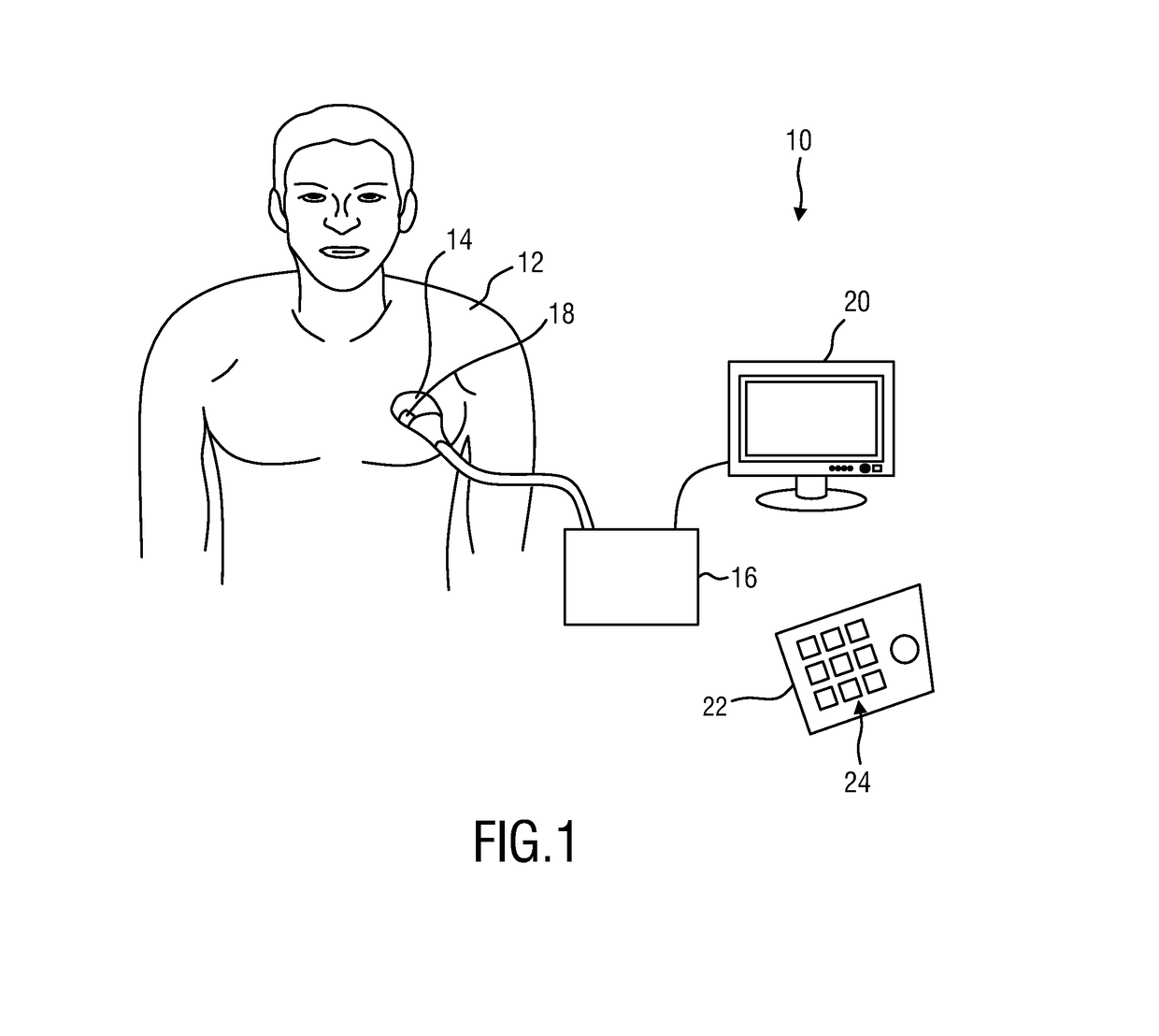
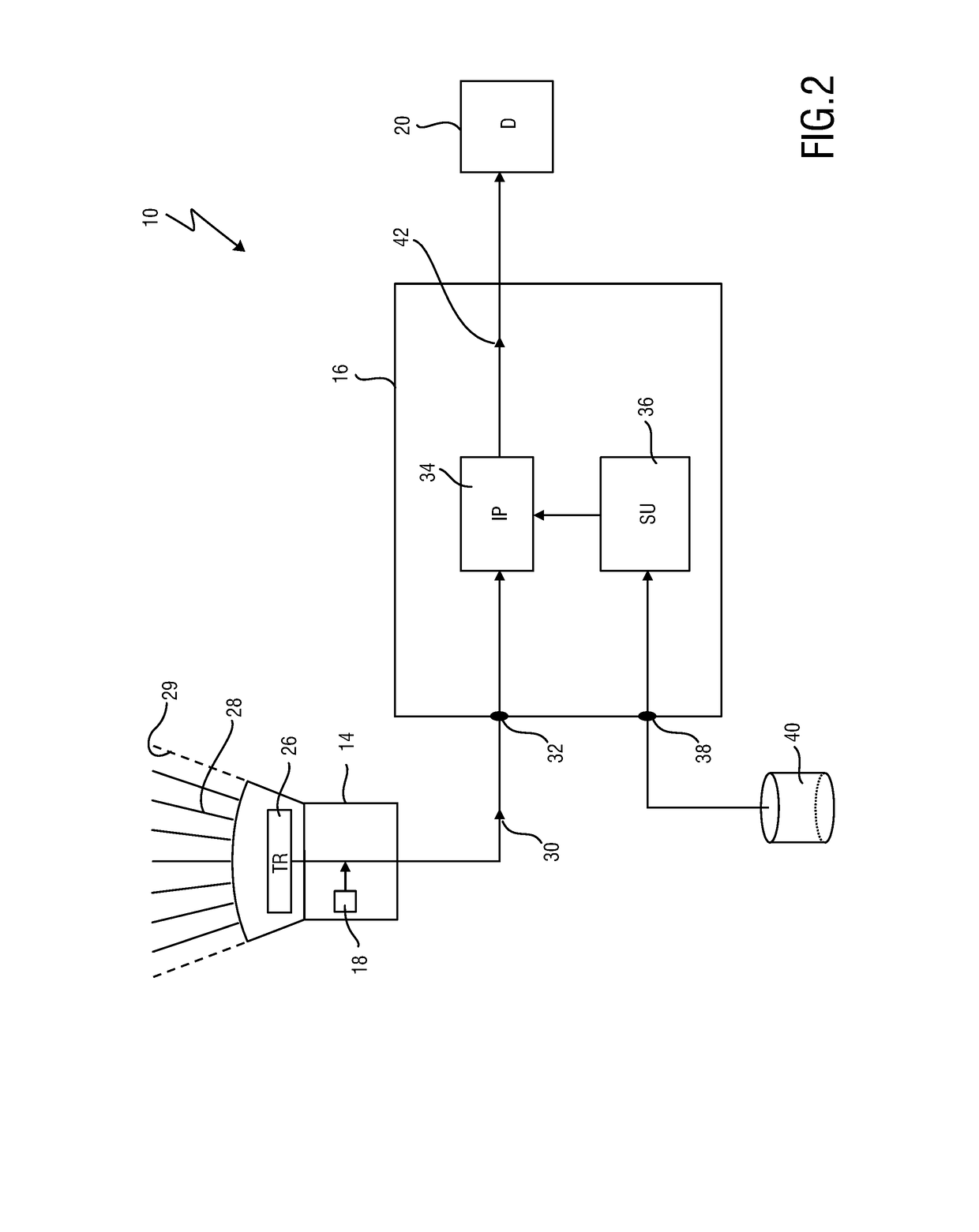
[0047]FIG. 1 shows a schematic illustration of an ultrasound imaging apparatus 10 according to an embodiment, in particular a medical ultrasound two-dimensional imaging system. The ultrasound imaging apparatus 10 is applied to inspect a volume of an anatomical site, in particular an anatomical site of a patient 12. The ultrasound imaging apparatus 10 comprises an ultrasound probe (or ultrasound acquisition unit) 14 having at least one transducer array having a multitude of transducer elements for transmitting and / or receiving ultrasound waves. The transducer elements are arranged in an array so that the ultrasound probe 14 can determine two-dimensional ultrasound data in a field of view in an image plane of the anatomical site of the patient 12.

[0048]The ultrasound imaging apparatus 10 comprises a control unit 16 that controls the ultrasound probe 14 and the acquisition of the ultrasound data. As will be explained in further detail below, the control unit 16 controls not only the ac...


PUM


Abstract

An ultrasound imaging apparatus (10) for segmenting an anatomical object in a field of view (29) of an ultrasound acquisition unit (14) is disclosed. The ultrasound imaging apparatus comprises a data interface (32) configured to receive a two-dimensional ultrasound data (30) of the object in the field of view in an image plane from the ultrasound acquisition unit and to receive a three-dimensional segmentation model (46) as a three-dimensional representation of the object from a segmentation unit (36). An image processor (34) is configured to determine a two-dimensional segmentation model (50) on the basis of the three-dimensional segmentation model and a segmentation plane (48), wherein the segmentation plane and an image plane of the two-dimensional ultrasound data correspond to each other. The image processor is configured to adapt a contour of the two-dimensional segmentation model to the two-dimensional ultrasound data on the basis of pattern detection and where the image processor is configured to provide annotated two-dimensional image data (42) on the basis of the two-dimensional ultrasound data and the adapted segmentation model aligned to the two-dimensional ultrasound data.

Description

FIELD OF THE INVENTION[0001]The present invention relates to an ultrasound imaging apparatus for segmenting anatomical objects in a field of view of an ultrasound acquisition unit. The present invention further relates to a method for segmenting an anatomical object in a field of view of an ultrasound acquisition unit. In particular, the present invention relates to image processing and segmentation of anatomical objects in a scanned volume of a patient. The present invention further relates to a computer program comprising program code means for causing a computer to carry out the steps of the method according to the invention when said computer program is carried out on a computer.BACKGROUND OF THE INVENTION[0002]In the field of medical imaging systems it is generally known to combine different images of a patient acquired by identical or different medical analysis systems in order to improve the diagnostic possibilities. In particular, ultrasound systems are known which combine u...

Claims


Application Information

Patent Timeline
IPC IPC(8): A61B8/14G06T7/12A61B8/08G06T7/149A61B8/00
CPCA61B8/14A61B8/5223G06T7/12G06T2207/30004A61B8/468G06T2200/04G06T2207/10132G06T7/149G06T2207/30244G06T2207/20128G06T7/0014G06T2207/10136G06T2219/004G06T7/75G16H50/30
Inventor PETERS, JOCHENTAHMASEBI MARAGHOOSH, AMIR MOHAMMADWEESE, JUERGENBUERGER, CHRISTIAN
Owner KONINKLJIJKE PHILIPS NV
Why Eureka
Try Eureka