
Disease model
- Summary
- Abstract
- Description
- Claims
- Application Information
AI Technical Summary
Benefits of technology
Problems solved by technology
Method used
Image

Examples
example
Example 1 Production of Lung Cancer Model
Method
1. Collection of Rat Lung
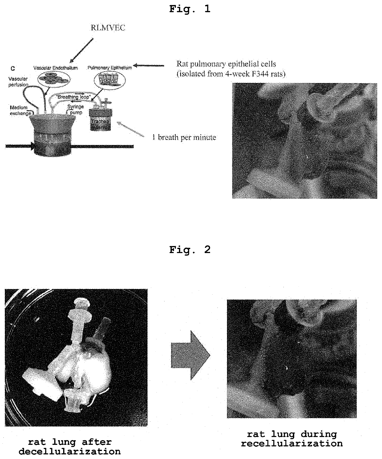
[0073]Lungs were collected from young adult (3 months old) male Fisher 344 rats (Charles River, Wilmington, Mass.). All animal experiments were conducted with the approval of the Institutional Animal Care and Use Committee of Nagasaki University, and in accordance with the animal experiment guidelines of Nagasaki University. Rats were euthanized by intraperitoneal injection of pentobarbital sodium (Sigma, 140 mg / kg) and heparin (250 U / kg). The diaphragm was punctured and the thorax was amputated to expose the lung. Lungs were perfused via the right ventricle with PBS containing 50 U / ml heparin (Sigma) and 1 μg / ml sodium nitroprusside (SNP, Fluka). After completion of the perfusion, the heart, lungs and trachea were dissected and removed all together. 18 G and 14 G catheters were respectively cannulated into the pulmonary artery, pulmonary vein and trachea.
2. Production of Lung Cancer Model Lung
2-1. Decellulariza...
example 2
Histological Analysis
Method
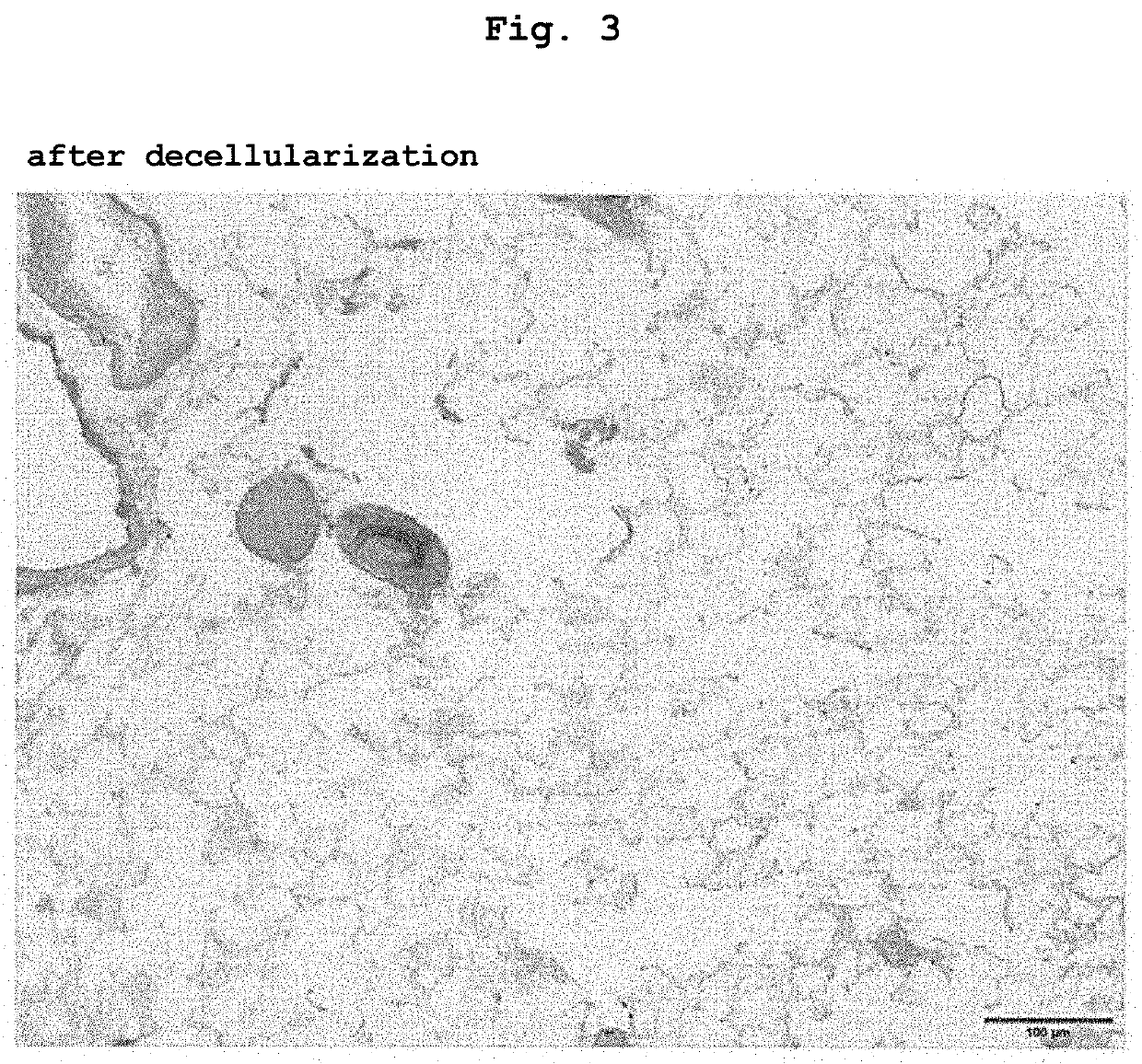
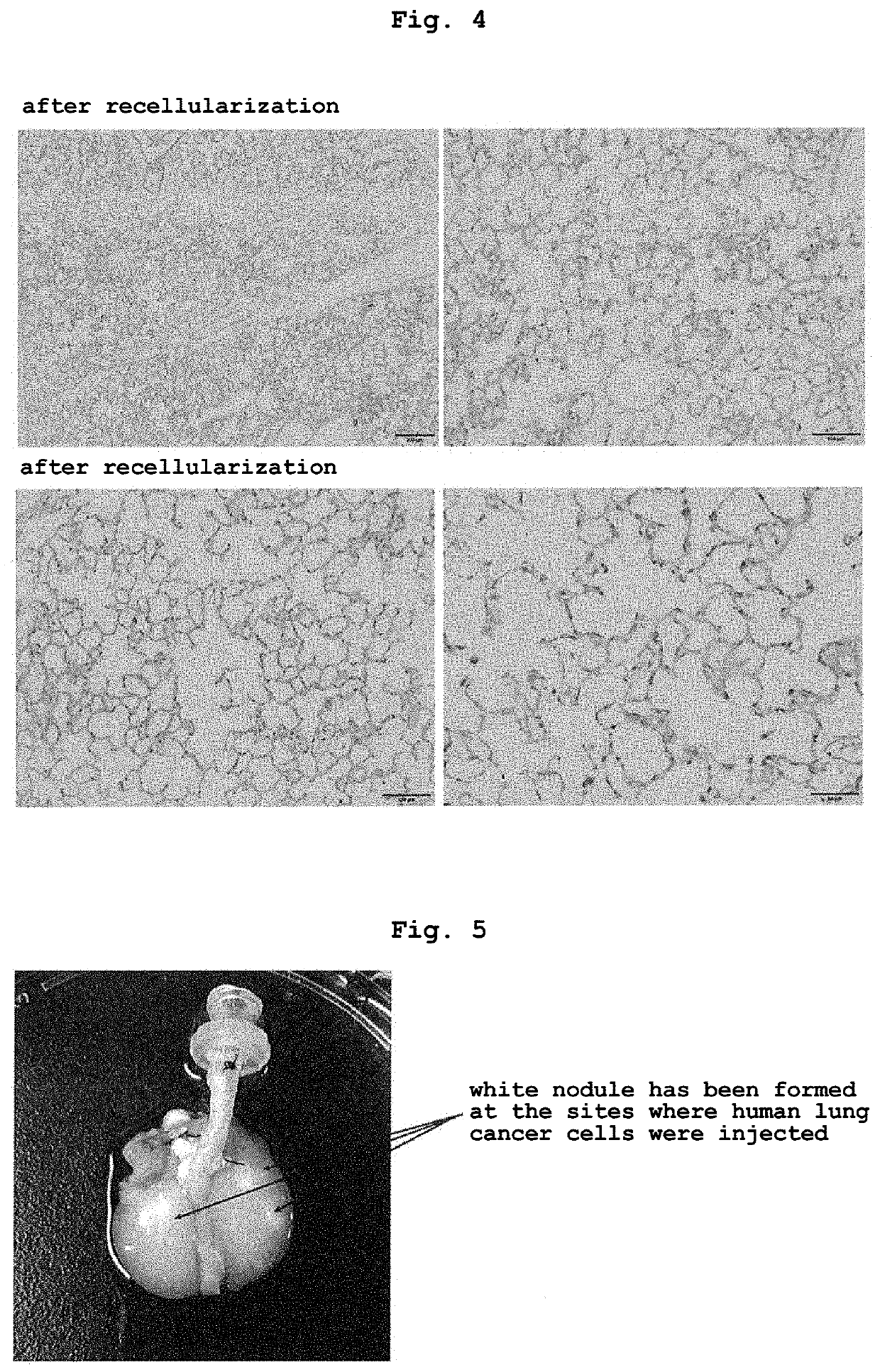
[0082]Samples (decellularized lung, recellularized lung or recellularized lung injected with cancer cell) were fixed for 4 hr in 10% formalin or 4% paraformaldehyde, dehydrated, embedded in paraffin, 5 μm sections were produced, and Hematoxylin-Eosin (H&E) staining was performed.
Periodic Acid-Schiff Staining
[0083]Samples (decellularized lung, recellularized lung or recellularized lung injected with cancer cell) were fixed for 4 hr in 10% formalin, dehydrated, embedded in paraffin, 5 μm sections were produced. Thereafter, the sections were deparaffinized and xylene was removed, and the sections were washed with water for several seconds, then immersed in 0.5% periodic acid solution for 10 min, washed with running water for 5 min, then immersed in distilled water for 2 min, and further immersed in Schiff's reagent for 15 min. Then, the sections were immersed in a sulfite solution for 2 min, 3 times, and then washed with running wate...
example 3
Histological Analysis (Immunostaining)
[0085]MUC-1 is expressed in many solid cancer cells. In particular, the C-terminal side is considered to be involved in the proliferation of cancer cells, promotion of infiltration ability, suppression of apoptosis, and the like through interaction with multiple molecules related to signal transduction. In general, the expression level of MUC-1 increases in cancer cells. MUC-1 is expressed with polarity on the cell surface in normal epithelial cells, but the polarity is considered to be lost in cancer cells (depolarized expression pattern). Using MUC-1 as an index, whether or not the lung cancer model produced by the production method of the present invention reproduces naturally-occurring lung cancer was verified.
Method
[0086]In immunostained samples (two-dimensionally cultured lung cancer cell line (2D), recellularized lung (3D) injected with cancer cells), 2D was fixed with 4% paraformaldehyde (PFA) for 10 min and 3D was fixed with 4% PFA for ...
PUM
 Abstract
Description
Claims
Application Information