Method and apparatus for time resolved optical imaging of biological tissues as part of animals

Active Publication Date: 2006-01-31
SOFTSCAN HEALTHCARE GROUP
View PDF6 Cites 40 Cited by
  • Summary
  • Abstract
  • Description
  • Claims
  • Application Information

AI Technical Summary

Benefits of technology

[0010]According to one broad aspect of the invention there is provided a system and method for time resolved optical imaging of biological tissues as part of an animal. The design of the optical components of the present system allows a beam of light to be directionally propagated through air, that is to say through free-space optics, to impinge on desired points of illuminations in a region of interest (ROI) of the tissue. Light re-emitted from the ti

Problems solved by technology

CW is the simplest and least expensive of the three techniques but provides only limited information with regards to the spatial distribution of internal optical attenuation of the object being imaged.
However, widespread utilization of optical imaging systems has been impeded by some undesirable characteristics of existing systems.
For example, optical imaging devices often require cumbersome arrangements of optical fibers that are used to transport the light to and from the object.
The type of arrangement for the optical components described in these references requires a time consuming alignment of the region of interest with t

Method used


Image

  • Method and apparatus for time resolved optical imaging of biological tissues as part of animals
  • Method and apparatus for time resolved optical imaging of biological tissues as part of animals
  • Method and apparatus for time resolved optical imaging of biological tissues as part of animals

Examples


Example

[0025]The invention relates to the field of optical imaging of turbid media such as biological tissues as parts of animals. While the following description of the preferred embodiment provides examples that relate to imaging of small mammals such as mice, it will be appreciated that the method can also be applied to larger animals and in particular to laboratory animals such as dogs, pigs and primates.

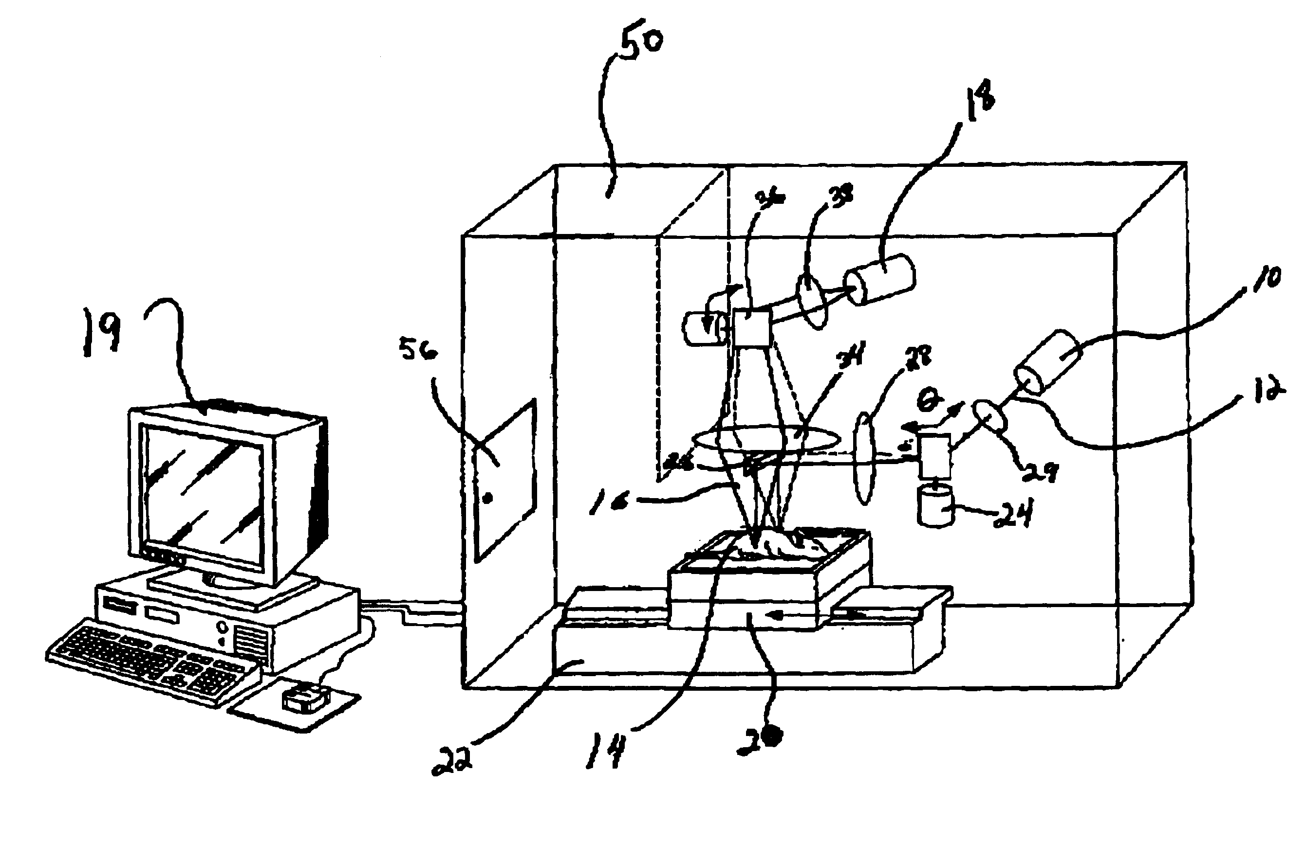
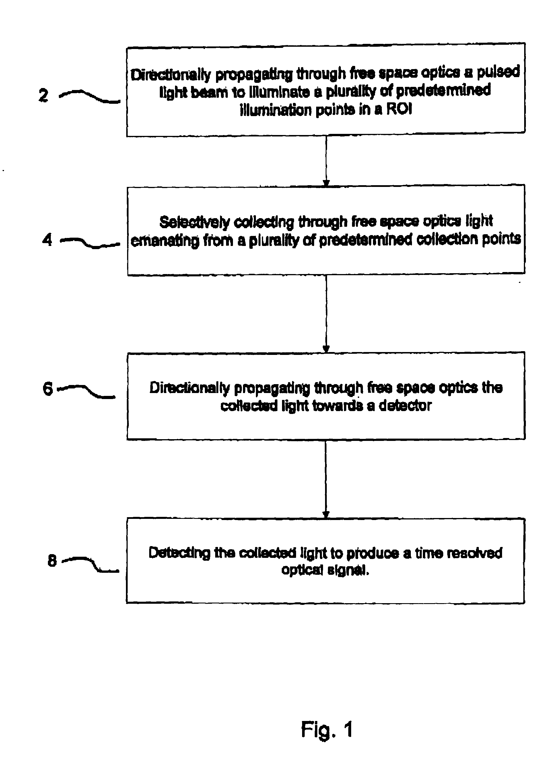
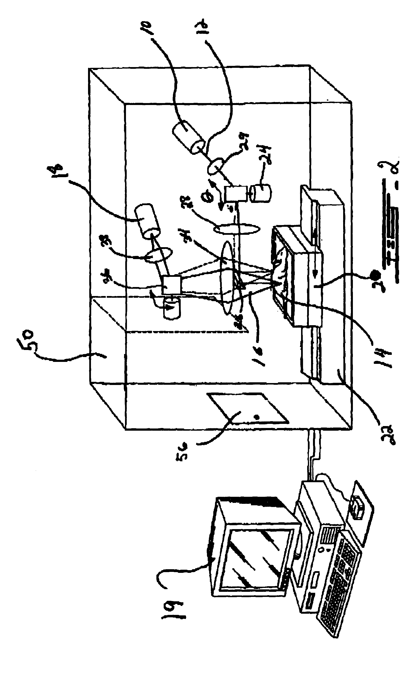
[0026]With reference to FIG. 1 an embodiment of the method of the present invention for collecting optical data for use in time resolved optical imaging is generally described. At 2 pulsed light from a source of a selected intensity is directionally propagated in air (i.e. through free space optics) to illuminate a plurality of predetermined illumination points in a ROI of comprising biological tissue within an animal. The light emanating from a plurality of collection points after diffusion through the tissue is selectively collected through free space optics at 4 and directionally pr...


PUM


Abstract

There is provided a method and system for collecting optical data for use in time resolved optical imaging wherein light is directionally propagated through free-space optics to impinge on a plurality of illumination at the surface of a biological tissue such as that comprised in small animals. Light re-emitted from the tissue is collected and directionally propagated through free space optics towards a detector to produce time resolved optical signals useful for optical image reconstructions.

Description

[0001]This application claims benefit of provisional application 60 / 505,352, filled Dec. 12, 2002CROSS-REFERENCE TO RELATED APPLICATIONS[0002]This application claims priority under 35USC§119(e) of U.S. patent application Ser. No. 10 / 317,856 filed Dec. 12, 2002 the petition to convert to a provisional patent application in accordance with 35USC§111(b)(6) and 37CFR1.53(c)(2) being filed concurrently herewith. This application is related to commonly assigned co-pending U.S. patent application Ser. No. 10 / 624,902 bearing agent docket number 15186-41US, the specification of which is hereby incorporated by reference.TECHNICAL FIELD[0003]The invention relates to the field of optical imaging of turbid media such as biological tissues as part of animals. More specifically, the invention relates to time resolved optical data acquisition for use in optical imaging.BACKGROUND OF THE INVENTION[0004]Different types of imaging techniques such as positron emission tomography (PET), magnetic resonan...

Claims


Application Information

Patent Timeline
IPC IPC(8): G01J3/30G01N21/64A61B5/00G01N21/47G01N21/49
CPCA61B5/0064A61B5/0073G01N21/6408G01N21/49G01N21/4795
Inventor LONG, WILLIAM F.BERUBE-LAUZIERE, YVESHALL, DAVID J.MCINTOSH, LAURA
Owner SOFTSCAN HEALTHCARE GROUP
Why Eureka
Try Eureka