Cervical fixation device

a cervical fixation and defect technology, applied in the field of cervical fixation or defect devices, can solve the problems of pain, deformity, stenosis of spinal canal or neuroforamina, and inability to fusion,

Inactive Publication Date: 2011-01-18
SPINAL ELEMENTS INC
View PDF79 Cites 80 Cited by
  • Summary
  • Abstract
  • Description
  • Claims
  • Application Information

AI Technical Summary

Problems solved by technology

Situations arise in which one or more cervical vertebrae 102 do not have adequate support or stability, which can lead to pain, deformity, stenosis of spinal canal or neuroforamina, and impairment or loss of nerve function.
However, loose bone fragments do not provide structural support and therefore fusion is often unsuccessful.
Structural bone grafts from the patient or donors have been successful, but may give rise to pain and complications if from the patient, and risk of disease transmission if from a donor.
Some cylindrical bone grafts and devices have also been associated with increased subsidence and kyphotic deformities, particularly in the cervical area.
Such devices usually have simple implanted metal markers in front and back to allow limited visualization of their position with x-rays or the like.
Such devices are made with thick, vertically straight walls to provide support strength, but once they subside a small amount the straight walls offer no effective resistance to excessive subsidence.
The surface area provided for fusion is also limited by the thick walls.
Polymer material in current use does not allow construction of sharp edges and fixation elements and does not allow for varied shapes which might solve many of the problems with subsidence.
In the cervical spine 102, it is known in the art that the addition of an anterior plate will limit subsidence and kyphotic deformity, but this adds cost and is not always successful.
Complications such as backing out of fixation screws and screw and plate breakage have been significant problems.
Anterior plates are difficult to install if more than two disk levels are fused, and may add to dysphagia.
If surgery is required at an adjacent level, it is almost always necessary to remove the plate to perform the surgery, which increases complexity and morbidity when further surgery is required.

Method used


Image

  • Cervical fixation device
  • Cervical fixation device
  • Cervical fixation device

Examples


Embodiment Construction

[0036]Certain terminology is used in the following description for convenience only and is not limiting. The words “right,”“left,”“lower” and “upper” designate directions in the drawings to which reference is made. The words “inwardly” and “outwardly” refer to directions toward and away from, respectively, the geometric center of a cervical fixation device in accordance with the present invention, and designated parts thereof. The terminology includes the words noted above, derivatives thereof and words of similar import. Unless specifically set forth herein, the terms “a”, “an” and “the” are not limited to one element but instead should be read as meaning “at least one”.

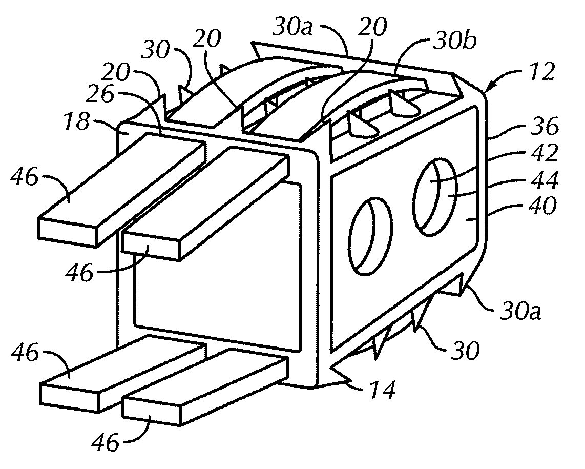
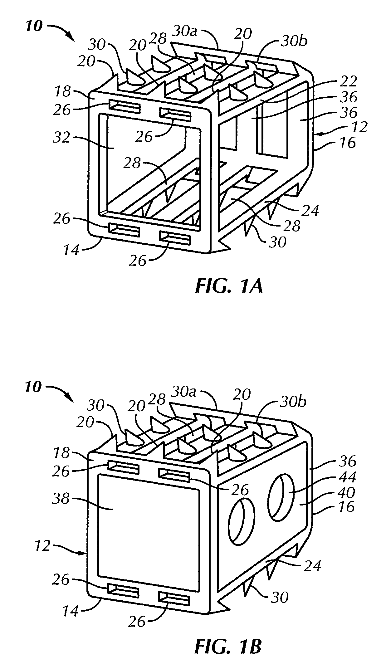
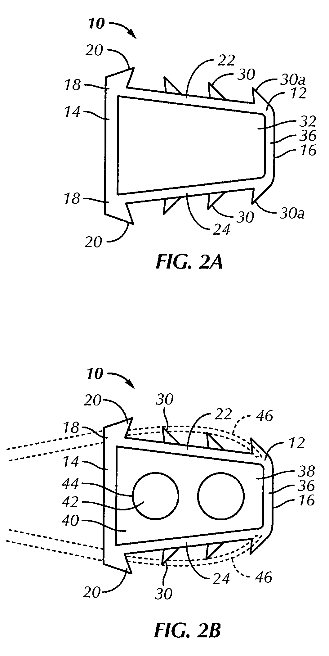
[0037]Referring to the drawings in detail, wherein like reference numerals indicate like elements throughout, there is shown in FIGS. 1A-5B a cervical fixation device, generally designated 10, 210, in accordance with first and second preferred embodiments of the present invention for insertion between a pair of adja...


PUM

PropertyMeasurementUnit
lengthaaaaaaaaaa
lengthaaaaaaaaaa
widthaaaaaaaaaa

Abstract

A cervical fixation device, for insertion between a pair of adjacent cervical vertebrae, includes a frame comprised of a first material. The frame has a generally rectangular proximal end and a generally rectangular distal end. The distal end is connected to the proximal end by at least one upper arch and at least one lower arch. The upper and lower arches are spaced apart from each other. The frame has a generally hollow interior, substantially open lateral sides between the upper and lower arches and a substantially open proximal end.

Description

CROSS-REFERENCE TO RELATED APPLICATIONS[0001]This application claims the benefit of U.S. Provisional Patent Application No. 60 / 745,895 filed Apr. 28, 2006 entitled “Dual Composition Vertebral Defect Device” which is incorporated by reference herein in its entirety.BACKGROUND OF THE INVENTION[0002]The present invention relates generally to vertebral fixation or defect devices, and more particularly to cervical fixation devices for insertion into an intervertebral space.[0003]As shown in prior art FIGS. 7A-11, it is known in the prior art that the spine 100, also known as the spinal column or vertebral column, supports the upper body, allows head, neck, and trunk motion, and includes twenty-four moveable vertebrae 101 including seven cervical vertebrae 102, twelve thoracic vertebrae 104, and five lumbar vertebrae 106, which extend from the skull to the sacrum.[0004]Referring to FIG. 8, with the exception of the first, upper most cervical vertebra 102, each vertebra 101 has a vertebral...

Claims


Application Information

Patent Timeline
Patent Type & Authority Patents(United States)
IPC IPC(8): A61F2/44
CPCA61F2/4455A61F2/28A61F2310/00359A61F2/447A61F2002/2835A61F2002/30056A61F2002/30062A61F2002/30112A61F2002/30331A61F2002/30433A61F2002/305A61F2002/30578A61F2002/30731A61F2002/30772A61F2002/30777A61F2002/30841A61F2002/30975A61F2002/4475A61F2002/4629A61F2210/0004A61F2220/0025A61F2220/0033A61F2220/0041A61F2230/0004A61F2250/0032A61F2310/00017A61F2310/00023A61F2310/00029A61F2310/00179A61F2002/30593
Inventor ECKMAN, WALTER W.
Owner SPINAL ELEMENTS INC
Why Eureka
Try Eureka