
A corneal biomechanical detection instrument and its application method
A biomechanics and testing instrument technology, applied in the field of corneal biomechanical testing instruments, can solve the problems of inability to take continuous pictures of corneal images, no difference in intensity, inaccurate testing results, etc., and achieve improved corneal biomechanical properties and corneal elasticity The effect of reducing, ensuring accuracy
Patent Information
- Authority / Receiving Office
- CN · China
- Patent Type
- Patents(China)
- Current Assignee / Owner
- Publication Date
- 2019-03-19
Smart Images
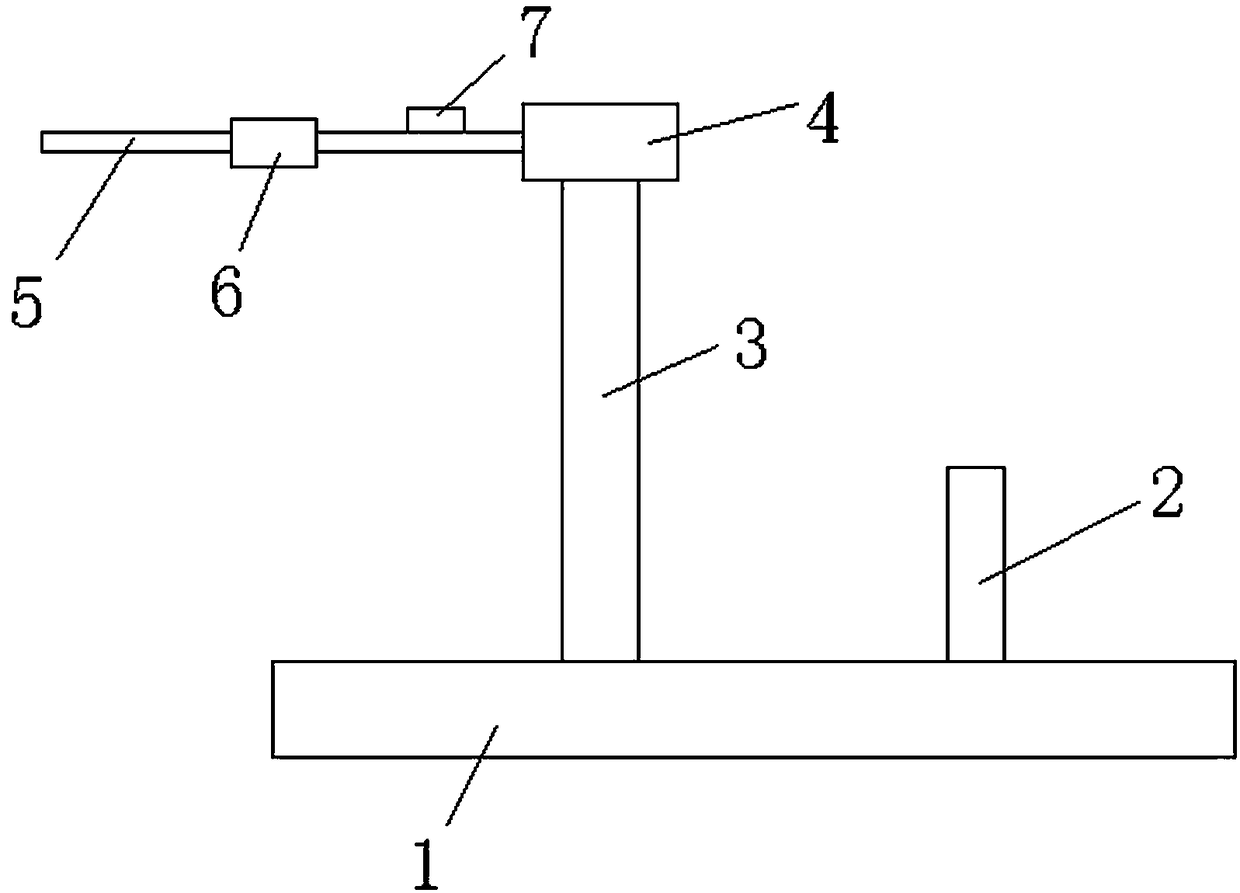
Figure 1 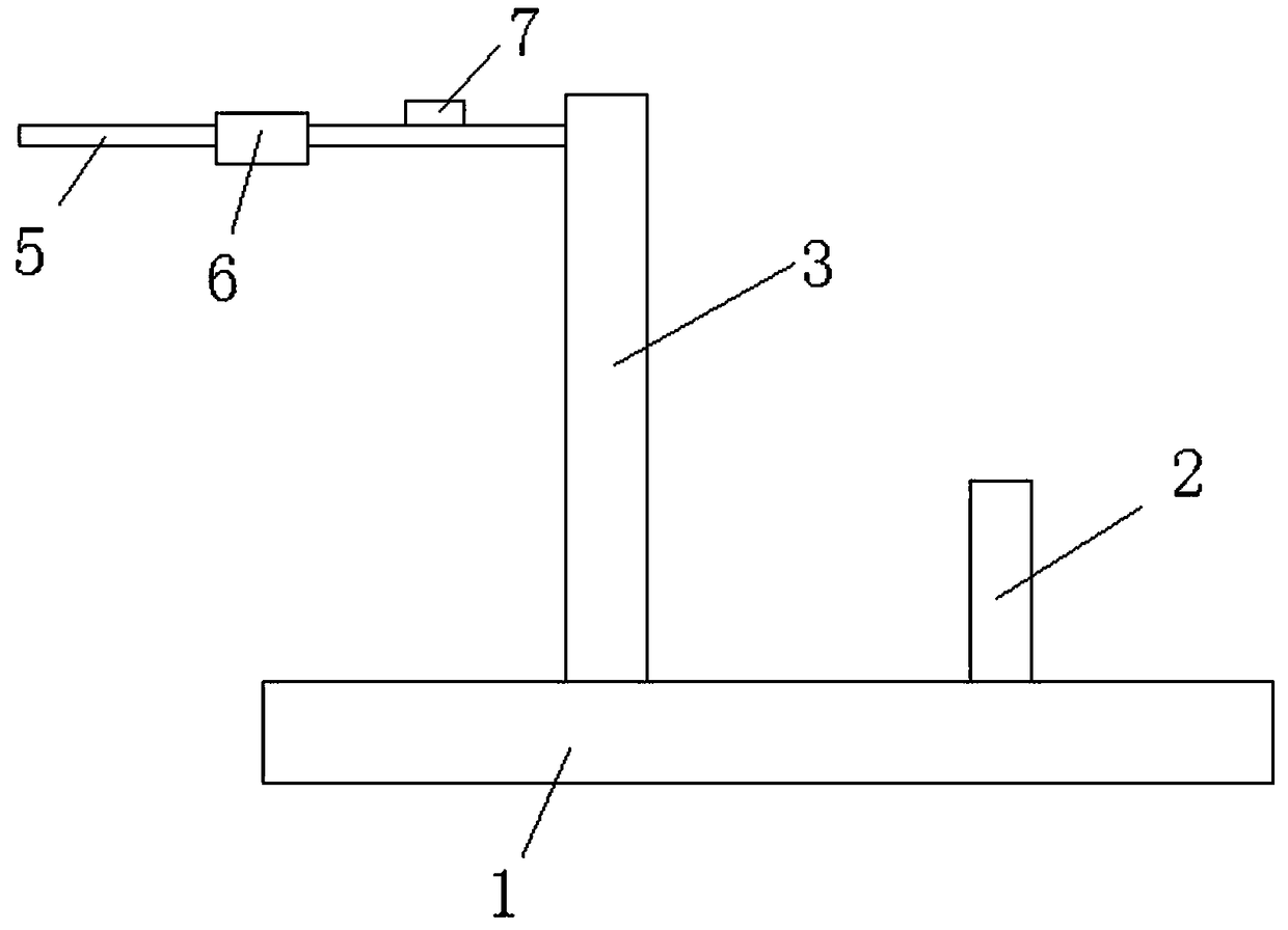
Figure 2 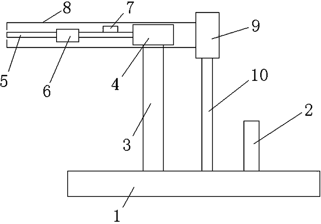
Figure 3
Abstract
Description
technical field
[0001] The invention belongs to the technical field of ophthalmology detection, and in particular relates to a corneal biomechanical detection instrument and a use method thereof. Background technique
[0002] The cornea is a complex biomechanical composite structure whose properties depend on structural subunits and patterns of assembly. In the cornea, only the Bowman's membrane and the stroma are collagen tissue layers, which are the main structural components that provide corneal strength; the corneal epithelium contributes little to the corneal strength, and the curvature of the cornea after corneal refractive surgery removes the corneal epithelium is almost No effect; the stretchability and low strength of the Descemet's membrane make it stretchable under a wide range of intraocular pressure, and it can well buffer and protect the corneal stroma that may cause damage to the corneal endothelium damage.
[0003] The structural material and mechanical pro...
Examples
Embodiment Construction
[0038] The technical scheme of the present invention will be described in detail below in conjunction with the accompanying drawings.
[0039] At present, the corneal elasticity of patients with keratoconus and corneal ectasia is reduced, which leads to the inability of the cornea to resist intraocular pressure, resulting in corneal deformation, changes in the refractive state of the eyeball, high myopia, and severe cases leading to corneal rupture and blindness. The existing non-invasive treatment of keratoconus and corneal ectasia is mainly corneal ultraviolet cross-linking, which can improve the biomechanical properties of the cornea, but so far there is no instrument that can effectively detect the improvement of corneal elasticity after ultraviolet cross-linking treatment .
[0040] The corneal biomechanics detection instrument and method provided by the present invention can detect the improvement of the corneal elasticity of the cornea treated by ultraviolet cross-linki...