
Apparatus and method to measure platelet contractility
a platelet and apparatus technology, applied in the field of apparatus and methods to measure platelet contractility, can solve the problems of insufficiently elucidating the cascading process, inability to completely understand the cascading process, and inability to induce clotting, so as to increase the scope of study, avoid operator exposure, and easy and inexpensive operation
Patent Information
- Authority / Receiving Office
- US · United States
- Patent Type
- Applications(United States)
- Current Assignee / Owner
- Publication Date
- 2009-06-11
- Estimated Expiration
- Not applicable · inactive patent
Smart Images
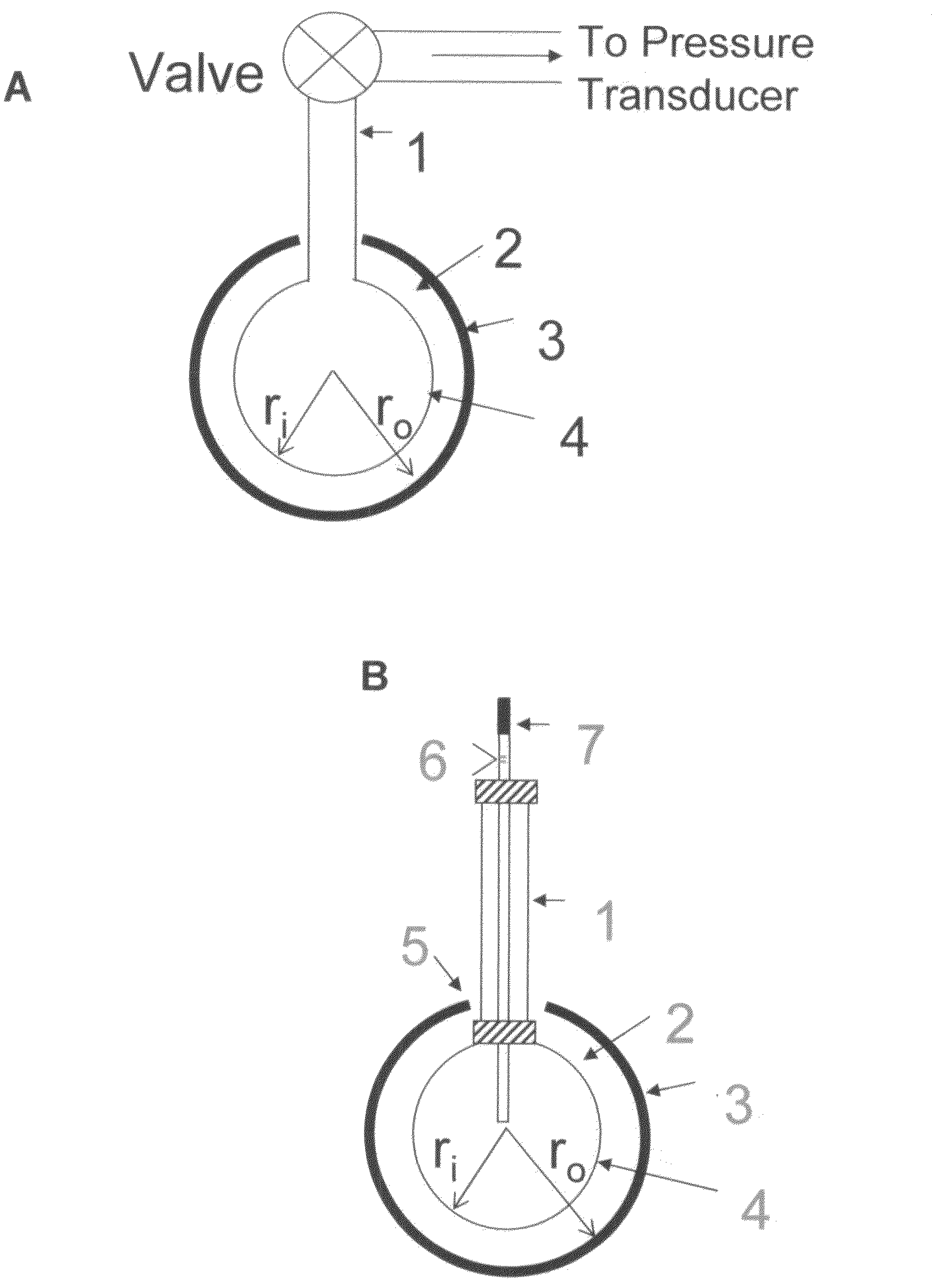
Figure 1 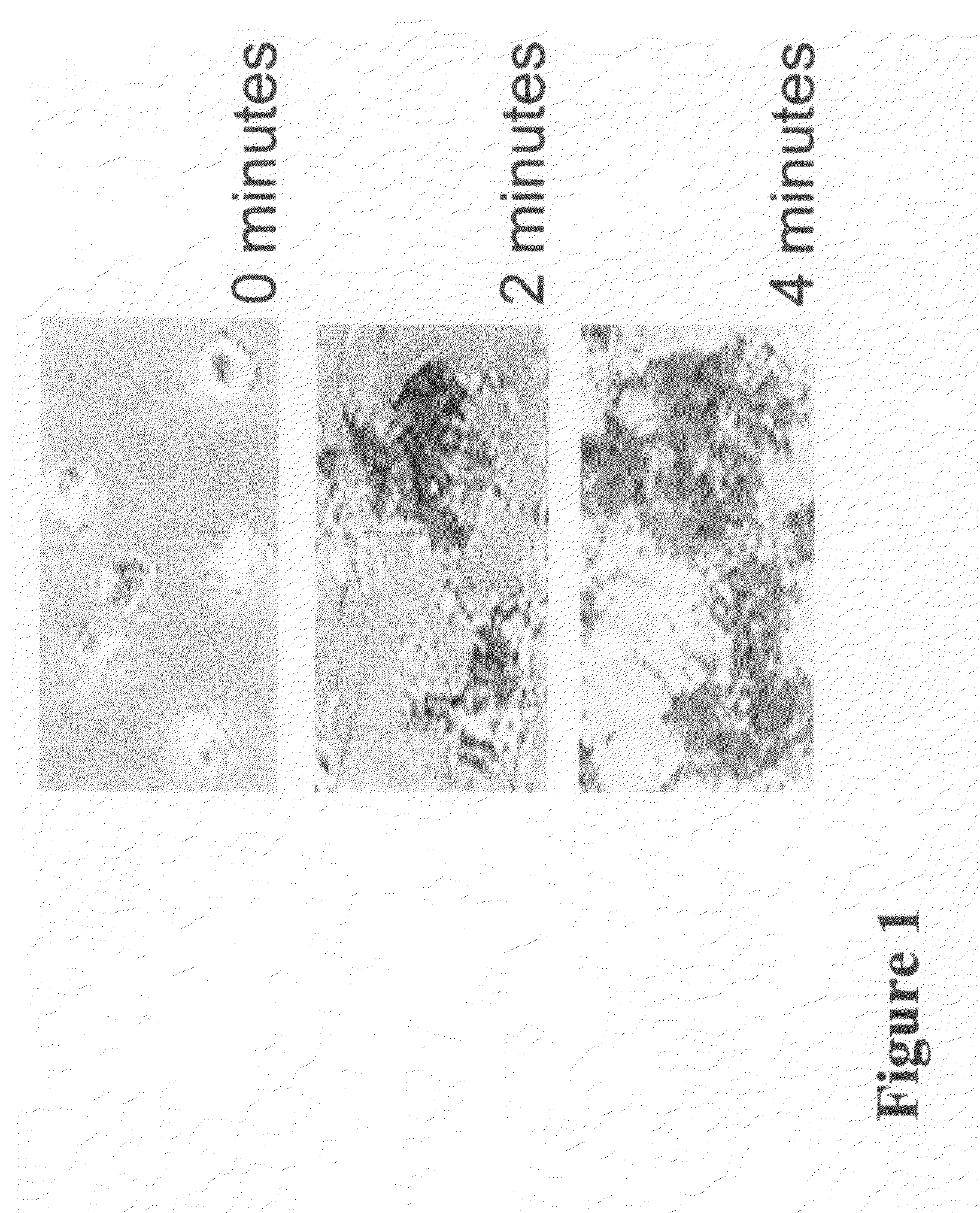
Figure 2 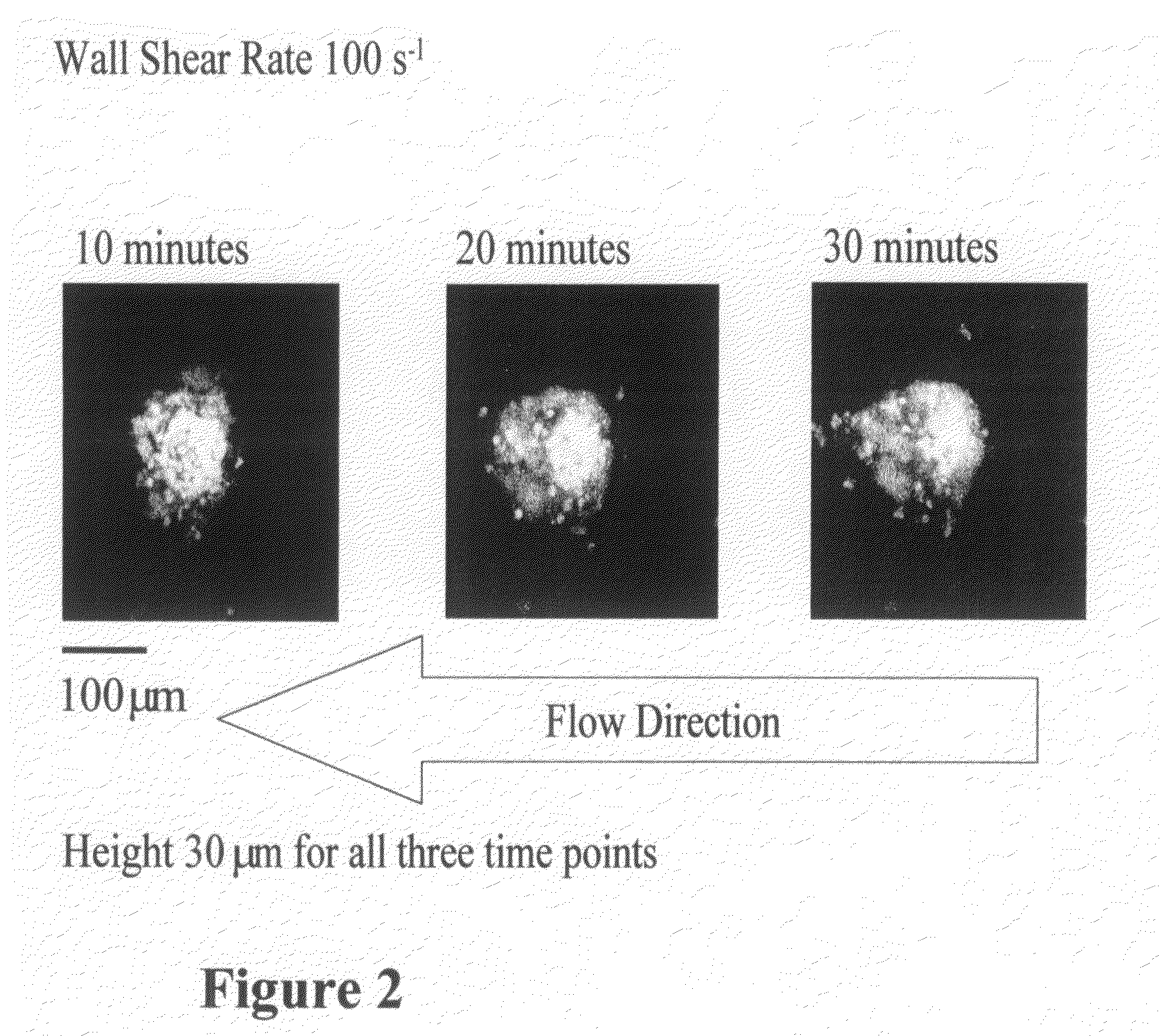
Figure 3
Abstract
Description
[0001] This application claims the benefit of priority under 35 U.S.C. § 121 of application Ser. No. 10 / 632,532, filed Aug. 1, 2003, which is incorporated by reference herein. This work was supported by National Institute of Health Grant No. R29-HL57430-01.BACKGROUND OF THE INVENTION
[0002] 1. Field of the Invention
[0003] The present invention generally concerns blood clotting components and mechanisms.
[0004] The present invention particularly concerns new devices and methods for studying clotting mechanisms and factors. More specifically, an apparatus and method for measuring and monitoring health and activity of platelets and other clotting factors are described. Most specifically, a clot retractometer and its method of use are provided to measure clot contractility forces as a means to provide a single point “funnel detection” procedure useful in aiding physiological and clinical research and patient diagnosis and monitoring of many diseases, as well as screening populations.
[0005] 2. ...
Examples
example 1
[0064]Perfusion studies.
[0065]Methods
[0066]Blood obtained from healthy volunteers was mixed with D-phenyl alanyl-L-prolyl-L-arginine chloromethyl ketone dihydrocloride (PPACK, 93: M) to prevent clotting. The platelet count was adjusted to 10,000 / μl to reduce the number of events on the surface and facilitate image analysis. Perfusion experiments were conducted in a parallel plate flow chamber at 37° C. (3) using type I collagen fibrils as reactive substrate onto glass coverslips. The interaction of flowing platelets with the surface was evaluated in real time by reflection interference contrast microscopy (RICM) using a Zeiss Axiovert 135M microscope. In this technique, interference colors indicate the distance between two surfaces, such as cellular membranes and a substrate coated on glass. On a gray scale, zero-order black indicates a separation of 4-12 nm, and white a distance >20-30 nm (15; 19). Experiments were recorded on S-VHS videotape at the rate of 30 frames per second and...
example 2
[0070]In order to demonstrate some of the technical capabilities currently being developed in the Inventors' laboratory, a summary of the development of a technique to create an upscale replica of an actual thrombus is presented below. The geometrical data of the thrombus are obtained with confocal microscopy while the blood is continuously flowing as previously reported (20). This technique was developed to study the flow field around a thrombus in an upscale chamber. By matching the Reynolds number, it is possible to determine the flow path in the microscopic realm, based on the similarity principle.
[0071]The evolution of an isolated thrombus at 100s−1 is depicted in FIG. 2. For the experiment shown here, a single thrombus was recorded from initiation of the flow. This image shows the growth changes in the thrombus once it is developed (>10 minutes) to 30 minutes. Each image is derived from the summation of a series of confocal image slices. Of note is the peculiar growth pattern ...
example 3
[0072]Below, Inventors describe their technical solution to create an upscale three-dimensional (3-D) model of a thrombus based on the information obtained with confocal microscopy.
[0073]As a first step, Inventors decided to investigate the already available techniques for 3-D rapid prototyping. A commonly used technique is stereolithography, which uses step-wise planar buildup of the object, based on the solidification of a photoresin by a laser beam. After each layer is cured, the object is lowered into a fluid resin pool by a distance equal to the vertical resolution of the system. This technique appears to be adequate in view of the complex geometries that it can handle.
[0074]The main practical difficulty in merging confocal microscopy and stereolithography is the lack of compatibility of the data. Confocal microscopy renders the data in a series of images (TIFF files in our system). These images are represented by a series of pixels with a given grayscale value, and the images ...