Micro-infusion method and device thereof based on epididymis duct intracavity environment experiment

A technology of epididymis and internal environment, applied in medical science, veterinary surgery, sensors, etc., can solve the problems of difficult fixation, epididymis and vas deferens intubation, avoid semen overflow, simple operation, avoid cutting vas deferens and Effects of cannula slippage

Inactive Publication Date: 2015-02-04
FOURTH MILITARY MEDICAL UNIVERSITY
View PDF7 Cites 2 Cited by
  • Summary
  • Abstract
  • Description
  • Claims
  • Application Information

AI Technical Summary

Problems solved by technology

[0003] The purpose of the present invention is to provide a microperfusion method and device based on the environment experiment in the epididymis lumen, so as to solve the problems of difficult intubation and fixation of the epididymis and vas deferens in the prior art

Method used


Image

  • Micro-infusion method and device thereof based on epididymis duct intracavity environment experiment

Examples


Embodiment Construction

[0026] The present invention will be described in detail below in combination with specific embodiments.

[0027] The microperfusion method based on the environment experiment of the epididymis lumen involved in the present invention is used for the in vivo study of the epididymis function of male animals, especially for the research of the epididymis epithelial function and the change of the microenvironment of the epididymis lumen. After exogenous intervention is applied to the lumen, the epididymis fluid can be collected to obtain information on changes after the intervention, and the normal epididymis lumen environment and physiological changes in epithelial cells can be observed. It is a method to obtain information as an intermediate result from a living body, specifically as follows Steps to achieve:

[0028] Step 1: intraperitoneally anesthetize the rat, and expose the epididymis through an incision near the middle of the scrotum;

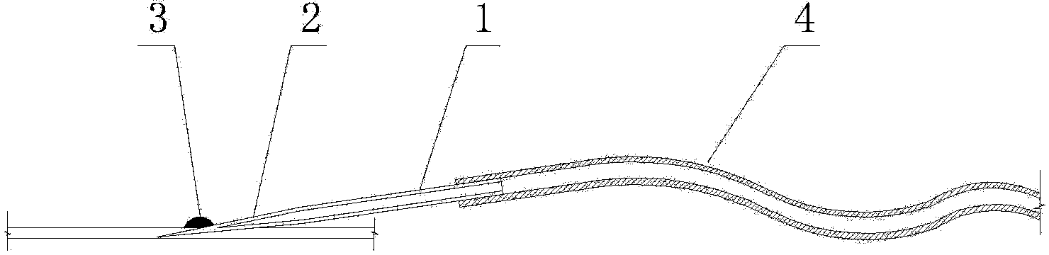
[0029] Step 2: Under a dissecting m...


PUM


Abstract

The invention relates to a micro-infusion method and a device thereof based on epididymis duct intracavity environment experiment. In international reports, the live micro-infusion method on the function research of epididymis ducts adopts the steps of by utilizing a dissecting microscope, separating the epididymis duct from the epididymis duct tail part, forming an incision, inserting a catheter, fixing by plasticine, separating an accompanying blood vessel near an spermaduct, and inserting a catheter, so the difficulty in intubation operation is larger. The method comprises the following steps of separating the epididymis duct from the epididymis duct tail part; using a glass micro-infusion tube, drawing and thinning a head part, and connecting the tail part with a silicone tube, a syringe and a micro injection pump; directly inserting a micro-infusion pipe head part into the epididymis duct, dripping a tissue glue for fixing, and filling interference materials; directly inserting the silicone tube without separation of the accompanying blood vessel near the spermaduct, and fixing by the tissue glue; finally, backflushing the spermaduct, and collecting epididymis duct liquid at the opening of the injection part of the epididymis duct. The method and the device have the advantages that the micro-infusion operation under the live environment is optimized, the operation is simple, the implementation is easy, the required material is purchased domestically, the experiment cycle is greatly shortened, and the experiment efficiency is improved.

Description

technical field [0001] The invention belongs to the field of epididymis intraluminal environment experiments, and in particular relates to a microperfusion method and a device based on the epididymis intraluminal environment experiment. Background technique [0002] At present, domestic research on the epididymis function of the reproductive system of male animals is mostly carried out in the state of tissue sampling and cell culture, but there are few reports on research in the living environment. Only a few research groups in the world use microperfusion in the living environment. To study the function of epididymis. They separated the epididymis duct from the tail of the epididymis, made an incision and intubated it, fixed it with plasticine, then made another opening at the proximal end of the vas deferens and intubated it, injected different substances into the tail of the epididymis by reverse perfusion, and studied the changes in the function of the epididymis. Howe...

Claims


Application Information

Patent Timeline
Patent Type & Authority Applications(China)
IPC IPC(8): A61D1/00
CPCA61D7/00A61B5/4387
Inventor 李臻马斌芳幸世龙程胖赵洁冯潇魏金花
Owner FOURTH MILITARY MEDICAL UNIVERSITY
Why Eureka
Try Eureka