
Blood cell analysis method and blood cell analyzer
A blood cell analysis, cell technology, applied in the direction of analysis of materials, material stimulation analysis, biological particle analysis, etc., can solve the problems of inaccurate platelet count, increase the cost of equipment and time, etc.
- Summary
- Abstract
- Description
- Claims
- Application Information
AI Technical Summary
Problems solved by technology
Method used
Image

Examples
Embodiment 1
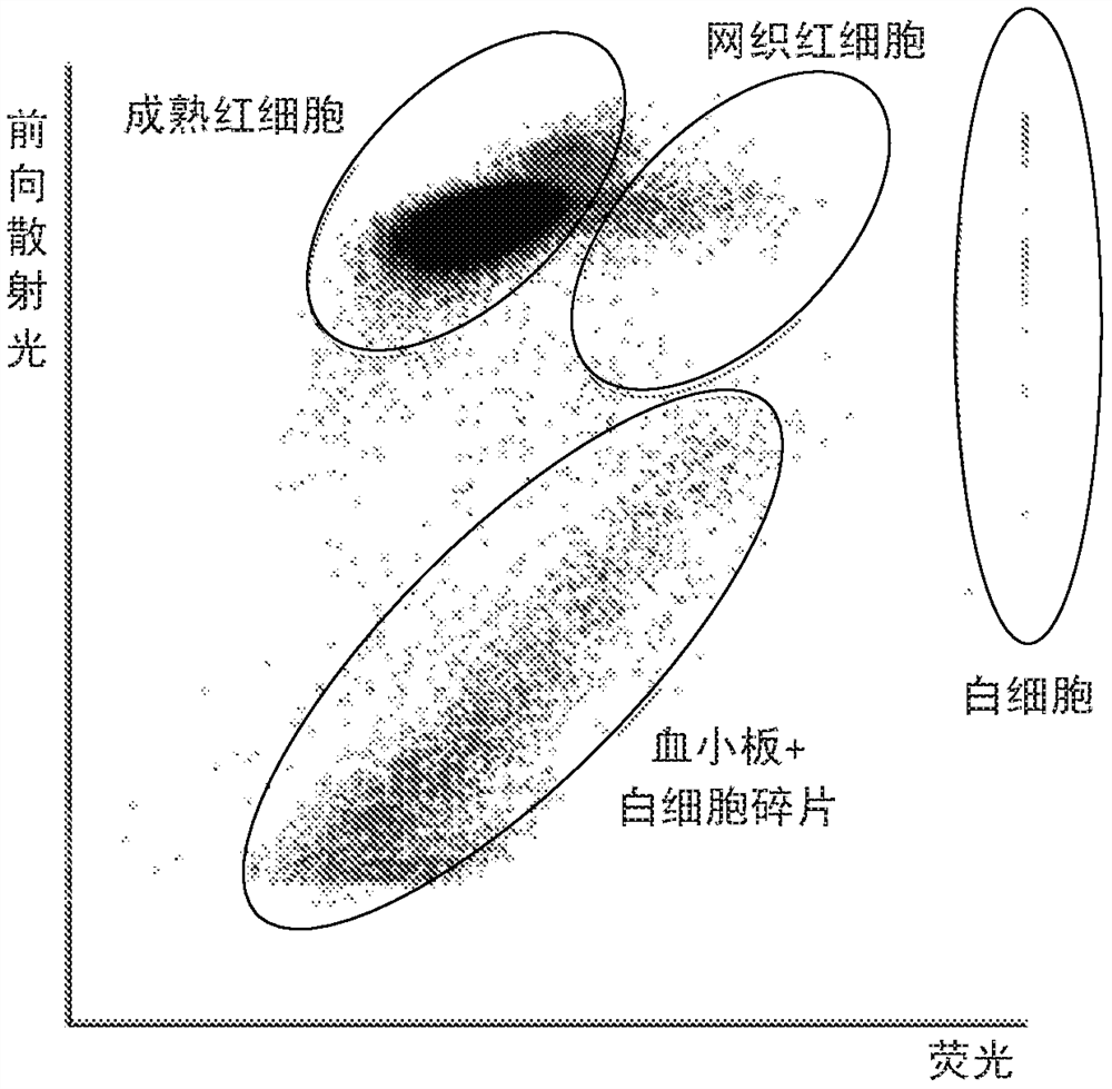
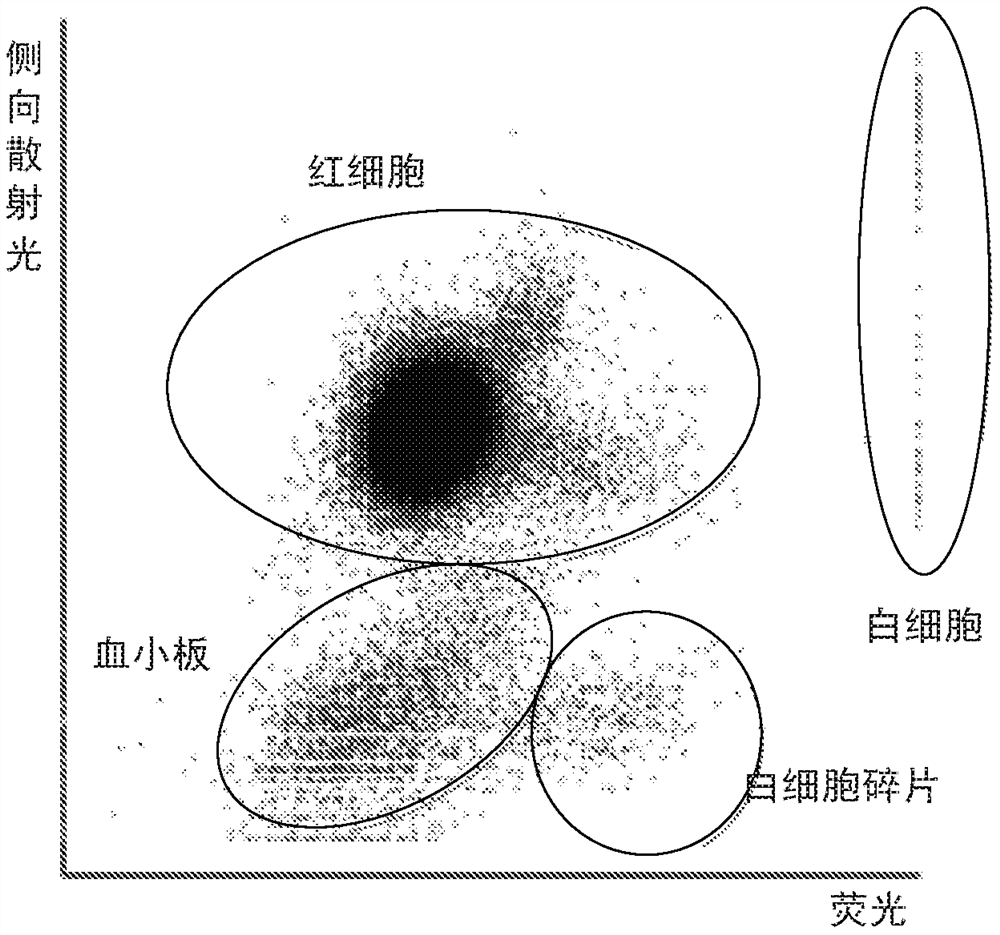
[0042] Provide the first blood sample containing white blood cell fragments, process the first blood sample to spheroidize the red blood cells, and basically maintain the integrity of the red blood cells, let the cells pass through the detection area of the blood cell analyzer one by one, and detect the precursor of each cell There are three signals: forward scattered light, side scattered light and fluorescence. in figure 2 showing the forward scattered light-fluorescence scattergram (first scattergram) of the first blood sample, image 3 A side scattered light-fluorescence scattergram (second scattergram) of the first blood sample is shown. figure 2 In , the distinction between platelets and white blood cell fragments is poor, so platelet counts cannot be obtained accurately. exist image 3 Among them, platelets and white blood cell fragments can be distinguished, but platelets and red blood cells are not well distinguished, so you can use image 3 Calculate the whit...
Embodiment 2
[0046] Provide a second blood sample containing white blood cell fragments, process the second blood sample to spheroidize red blood cells and maintain the integrity of red blood cells, let the cells pass through the detection area of the blood cell analyzer one by one, and detect the forward direction of each cell Scattered light, side scattered light and fluorescence three signals. in Figure 5 shows the forward scattered light-fluorescence scatter diagram of the second blood sample, Image 6 A side scatter-fluorescence scatter plot of the second blood sample is shown. Figure 5 In , the distinction between platelets and white blood cell fragments is poor, so platelet counts cannot be obtained accurately. exist Image 6 In , platelets and white blood cell fragments can be distinguished, but platelets and red blood cells are not distinguished, so they can be distinguished in Image 6 Determine the preset feature area in the preset feature area, and identify the scattere...
PUM
 Abstract
Description
Claims
Application Information