

Coupling method of fluorescent latex microspheres and protein
A fluorescent latex microsphere and coupling technology, applied in the field of biological analysis, can solve the problems of low coupling efficiency and coupling strength, low fluorescence value of the detection card, etc., to improve stability, avoid agglutination, and improve coupling efficiency and the effect of coupling strength
- Summary
- Abstract
- Description
- Claims
- Application Information
AI Technical Summary
Problems solved by technology
Method used
Image

Examples
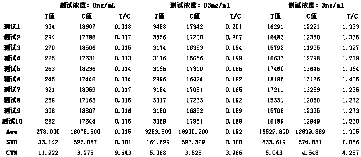
Embodiment 1
[0029] The present embodiment is a coupling method of fluorescent latex microspheres and cardiac troponin I monoclonal antibody, and the coupling method comprises the following steps:
[0030] (a) Dilute the fluorescent latex microspheres with MES buffer to a solid content of 0.8‰; then add an ethanol solution containing NHS and an ethanol solution containing EDC to the diluted fluorescent latex microsphere solution, mix well, and react at room temperature 25min, sonication, centrifugation at 10000r / min for 10min, collecting and collecting the activated fluorescent latex microspheres, adding MES buffer, ultrasonic treatment to obtain a fluorescent latex microsphere solution with a solid content of 0.08% of the fluorescent latex microspheres, wherein, The volume ratio of the volume ratio of the ethanol solution containing NHS and the ethanol solution containing EDC to the fluorescent latex microsphere solution is 1:26; the concentration of NHS in the ethanol solution containing ...
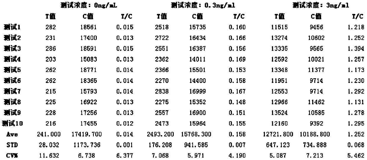
Embodiment 2
[0035] The present embodiment is a coupling method of fluorescent latex microspheres and cardiac troponin I monoclonal antibody, and the coupling method comprises the following steps:
[0036] (a) Dilute the fluorescent latex microspheres with MES buffer to a solid content of 0.8‰; then add an ethanol solution containing NHS and an ethanol solution containing EDC to the diluted fluorescent latex microsphere solution, mix well, and react at room temperature 25min, sonication, centrifugation at 10000r / min for 10min, collecting and collecting the activated fluorescent latex microspheres, adding MES buffer, ultrasonic treatment to obtain a fluorescent latex microsphere solution with a solid content of 0.08% of the fluorescent latex microspheres, wherein, The volume ratio of the volume ratio of the ethanol solution containing NHS and the ethanol solution containing EDC to the fluorescent latex microsphere solution is 1:28; the concentration of NHS in the ethanol solution containing ...
Embodiment 3
[0041] The present embodiment is a coupling method of fluorescent latex microspheres and cardiac troponin I monoclonal antibody, and the coupling method comprises the following steps:
[0042](a) Dilute the fluorescent latex microspheres with MES buffer to a solid content of 1.2‰; then add an ethanol solution containing NHS and an ethanol solution containing EDC to the diluted fluorescent latex microsphere solution, mix well, and react at room temperature 25min, sonication, centrifugation at 10000r / min for 10min, collecting and collecting the activated fluorescent latex microspheres, adding MES buffer, ultrasonic treatment to obtain a fluorescent latex microsphere solution with a solid content of 0.08% of the fluorescent latex microspheres, wherein, The volume ratio of the volume ratio of the ethanol solution containing NHS and the ethanol solution containing EDC to the fluorescent latex microsphere solution is 1:22; the concentration of NHS in the ethanol solution containing N...
PUM
 Abstract
Description
Claims
Application Information